CT vs MRI: Key Differences & When to Use
The Question Every Student Asks
"Should this patient get a CT or an MRI?" It's the most common imaging decision in modern medicine, and the answer is rarely straightforward. Both are cross-sectional modalities that revolutionized diagnostic imaging — but they work on completely different physical principles and excel at different things.
TL;DR — A Safe Starting Point
CT is usually faster and is strong for cortical bone, lungs, acute hemorrhage, and many emergency protocols. It uses ionizing radiation.
MRI offers versatile soft-tissue contrast without ionizing radiation. It usually takes longer and requires rigorous screening for implants, foreign bodies, and other hazards. The correct choice depends on the indication, protocol, urgency, and patient.
How They Work — The Physics Simplified
CT (Computed Tomography)
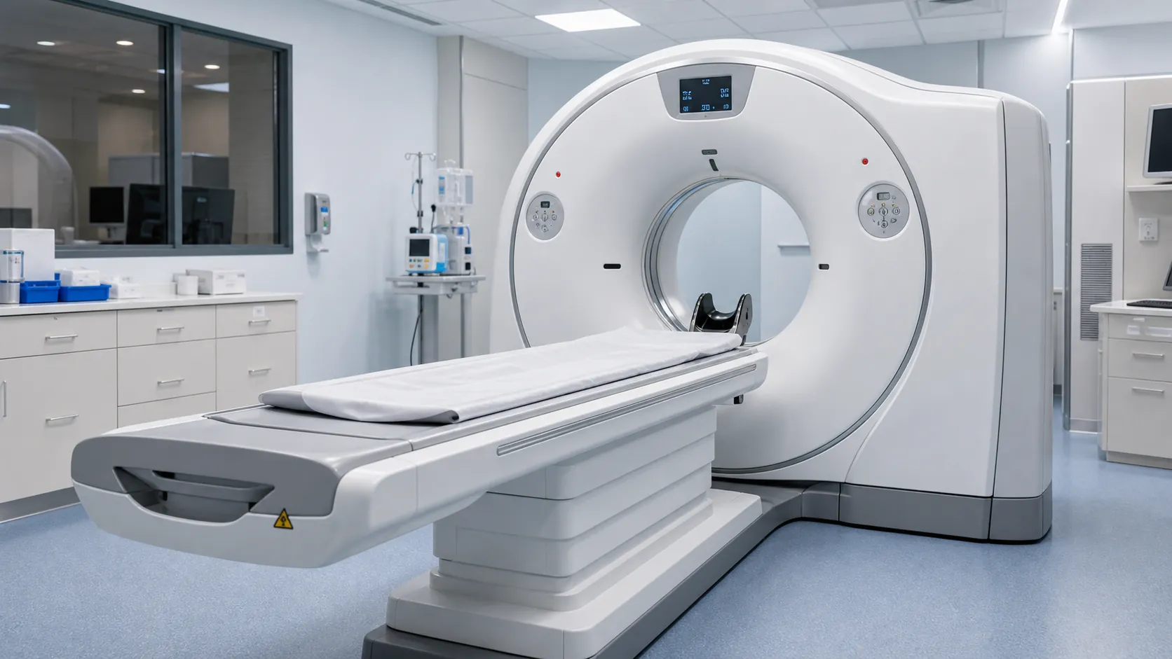
A CT scanner rotates an X-ray tube and detector array around the patient, acquiring projections from different angles. A computer reconstructs cross-sectional images using methods such as filtered back projection or iterative reconstruction. Pixel values are reported in Hounsfield units (HU), an attenuation scale referenced to water (0 HU); air is approximately −1000 HU. Bone and metal span broad, scanner- and material-dependent positive values, so fixed ranges should not be treated as universal.
Key traits: High spatial resolution and acquisition measured in seconds for many protocols; particularly useful for cortical bone, lung, calcification, and acute hemorrhage. CT uses ionizing radiation. Effective dose varies greatly with body region, patient size, scanner, and protocol—for example, RadiologyInfo lists approximate adult values of 1.6 mSv for a brain CT, 6.1 mSv for chest CT, and 7.7 mSv for abdomen/pelvis CT, versus 0.1 mSv for a two-view chest radiograph. These population-level estimates are not individual dose measurements.
MRI (Magnetic Resonance Imaging)
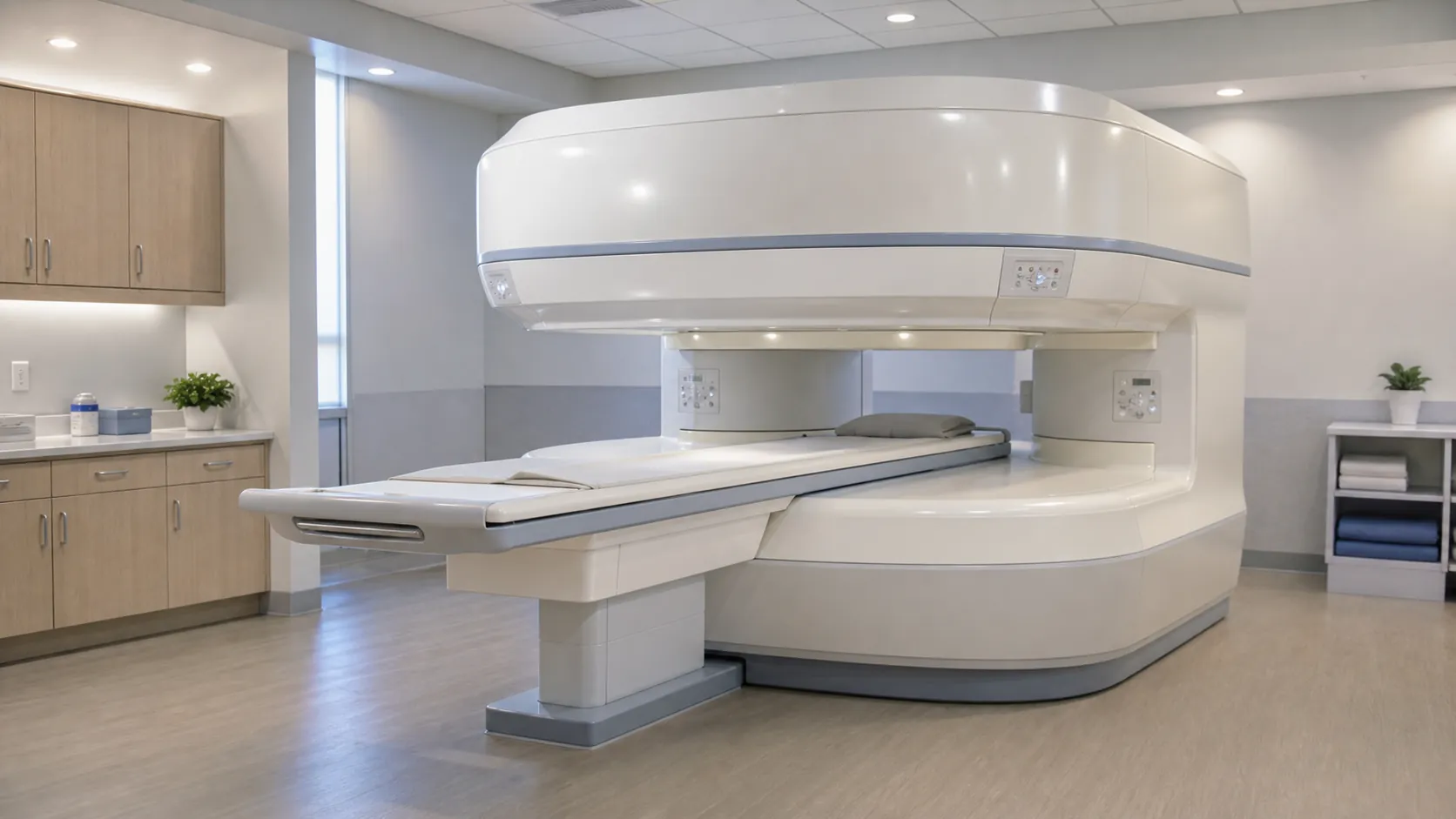
MRI uses a strong static magnetic field, switched gradient fields, and radiofrequency (RF) energy; it does not use ionizing radiation. Clinical systems commonly operate at 1.5 T or 3 T, although other field strengths exist. Hydrogen nuclei generate signals whose T1 and T2 relaxation, proton-density, diffusion, flow, and other properties can be weighted to produce different tissue contrasts.
Key traits: Versatile soft-tissue contrast and multiplanar imaging without ionizing radiation. Examinations are usually longer than CT and can be loud and motion-sensitive. Implants are not a single blanket contraindication: each device must be positively identified and scanned only under its documented MR safety conditions or an institutionally approved expert protocol.
Head-to-Head: Where Each Excels
| Clinical Question | Commonly appropriate approach | Important qualification |
|---|---|---|
| Acute head trauma | Usually noncontrast head CT | Rapidly evaluates clinically important acute hemorrhage and fractures. MRI is useful when neurologic findings persist despite an unrevealing CT or when CT does not explain the deficits. |
| Suspected acute stroke | Emergency stroke protocol | Noncontrast CT is commonly used first to exclude hemorrhage; CTA evaluates large-vessel occlusion and CTA/CT perfusion may guide treatment in selected patients. MRI with diffusion is highly sensitive for acute ischemia and is appropriate when immediately available without treatment delay. The protocol depends on onset, treatment pathway, and local resources. |
| Brain tumor characterization | Usually MRI without and with contrast | MRI generally best defines tumor extent and tissue characteristics. CT remains useful for calcification, bone involvement, emergencies, or when MRI cannot be performed; MR spectroscopy is an optional problem-solving technique, not routine in every case. |
| Multiple sclerosis | MRI | Standardized brain and, when indicated, spinal cord MRI protocols assess lesions and disease activity. CT is not the preferred test for demyelinating lesions. |
| Spinal trauma / suspected fracture | CT for patients who meet imaging criteria | CT is generally first-line in high-risk blunt cervical spine trauma. MRI is added for suspected cord, ligamentous, or nerve-root injury and selected unexplained neurologic deficits; imaging is not automatically required for every low-risk patient. |
| Spinal cord compression | Usually MRI | MRI directly evaluates the cord, canal, discs, infection, and tumor. CT myelography can be used when MRI is contraindicated or nondiagnostic. |
| Suspected pulmonary embolism | Usually CTPA after clinical risk assessment | CTPA is commonly appropriate when PE is sufficiently likely or D-dimer is positive. Ventilation/perfusion imaging is an important alternative in selected patients, including some with iodinated-contrast contraindications or during pregnancy. |
| Chest / lungs | Usually CT for lung parenchyma, nodules, and trauma | MRI is not a general replacement for chest CT because lung signal and motion can limit it, but MRI has selected uses for mediastinal, pleural, chest-wall, vascular, and pediatric questions. |
| Liver lesion characterization | Multiphasic CT or MRI, depending on the lesion | MRI, sometimes with a hepatobiliary agent, can add tissue characterization; multiphasic CT is also appropriate for many lesions and oncology protocols. Ultrasound or contrast-enhanced ultrasound may be appropriate in selected settings. |
| Suspected urinary stones | Low-dose noncontrast CT for many nonpregnant adults | Ultrasound is generally preferred first during pregnancy and commonly in children to avoid radiation. CT is highly accurate, but sensitivity varies with dose, size, and composition; “radiopaque” is an x-ray descriptor and is not why CT detects most stones. |
| Knee internal derangement | Usually MRI after radiographs when indicated | MRI assesses menisci, ligaments, cartilage, and marrow. CT or CT arthrography has selected roles when MRI is unavailable or contraindicated. |
| Shoulder rotator cuff | Usually ultrasound or MRI after radiographs | Choice depends on expertise and whether labral or deeper structural assessment is needed. CT arthrography can be appropriate when MR arthrography cannot be performed; therefore CT is not categorically “inappropriate.” |
| Cardiac imaging | Question-specific CT or MRI | Coronary CT angiography evaluates coronary anatomy; cardiac MRI is strong for ventricular function, tissue characterization, inflammation, viability, and congenital disease. Heart rate, rhythm, devices, renal function, and local expertise influence selection. |
| Oncology | Question- and tumor-specific | CT is widely used for chest/abdomen/pelvis staging and response assessment; MRI is preferred for many brain, liver, pelvic, marrow, and local soft-tissue questions. PET/CT and other modalities may be required. Neither CT nor MRI is universally best for “cancer.” |
Safety, Screening, and Contrast
CT and Iodinated Contrast
- Pregnancy is not an automatic CT contraindication. Ultrasound and MRI (usually without gadolinium) are preferred when they can answer the question, but ACOG states that needed radiography or CT generally should not be withheld because diagnostic exposures are usually far below doses associated with fetal harm. Justify and optimize the examination, especially when the fetus is in or near the scan range.
- Prior contrast reaction. A prior allergic-like reaction to the same class of contrast is the strongest predictor of another reaction. This is not an “iodine allergy,” and shellfish or povidone-iodine allergy alone is not a special risk factor. Depending on reaction severity and clinical need, options include another test, noncontrast CT, changing agent, or premedication; premedication does not prevent every reaction.
- Kidney function. Do not label all post-contrast AKI “contrast-induced nephropathy.” ACR distinguishes contrast-associated AKI (temporal association) from contrast-induced AKI (caused by contrast). Modern evidence suggests IV iodinated contrast is rarely independently nephrotoxic when baseline eGFR is stable and at least 30 mL/min/1.73 m²; patients with acute kidney injury or eGFR below 30 need an individualized risk-benefit assessment and preventive strategy when appropriate.
- Thyroid disease. Iodinated contrast can rarely precipitate thyrotoxicosis, but ACR does not recommend restriction solely for a history of hyperthyroidism. Avoid it in active thyroid storm when possible, and consider interference with planned radioactive-iodine imaging or therapy.
MRI, Implants, and Gadolinium Contrast
- Implants and devices. Screen every patient and accompanying person. Pacemakers, defibrillators, aneurysm clips, cochlear implants, neurostimulators, pumps, and other devices must be identified and checked against manufacturer conditions. “MR Conditional” means safe only under specified conditions; “non-MR-conditional” is not synonymous with an absolute ban, because some cardiac devices can be scanned at experienced centers under strict multidisciplinary protocols.
- Aneurysm clips and metallic foreign bodies. Do not scan until an intracranial aneurysm clip is documented as MR Conditional or MR Safe for the proposed conditions. Suspected orbital metal requires a risk history and, when indicated, prior radiographs or review of adequate CT—not automatic orbital radiographs for everyone. An unsafe ferromagnetic object can move, heat, malfunction, or cause major artifact.
- Claustrophobia, size, noise, and motion. These are limitations rather than absolute contraindications. Coaching, wider-bore equipment, protocol modification, anxiolysis, or monitored sedation may help; sedation adds its own staffing and physiologic risks.
- Gadolinium and kidneys. Gadolinium-based contrast agents (GBCAs) can cause acute reactions and are retained in trace amounts. Nephrogenic systemic fibrosis (NSF) is associated mainly with certain GBCAs in severe kidney dysfunction. ACR considers NSF risk with standard-dose Group II agents sufficiently low that renal-function assessment is optional before Group II administration, although contrast should still be necessary and the agent and dose selected appropriately.
- Gadolinium in pregnancy. Use a GBCA only when it is expected to provide critical benefit that outweighs potential fetal risk. ACOG advises limiting gadolinium during pregnancy; MRI itself does not use ionizing radiation. Breastfeeding ordinarily does not need to be interrupted after iodinated contrast or a GBCA.
Pediatric Principle
Children are more radiosensitive and have a longer lifetime in which radiation effects could appear. Use ultrasound or MRI when diagnostically appropriate, and when CT is the right test, tailor technique to child size and the clinical question. MRI may avoid radiation but can require longer immobility or sedation; that tradeoff must be considered rather than assuming MRI is always safer overall.
Cost, Time, and Availability — The Real-World Factors
CT image acquisition usually takes seconds to a few minutes, while many MRI acquisitions take substantially longer. Total room or visit time for either modality may increase with patient preparation, multiphase or specialized protocols, contrast, monitoring, reconstruction, or sedation. CT is generally more available and less expensive than MRI, but unsupported national price ranges are misleading: patient cost and scheduling vary by examination, facility, region, payer, and care setting.
In emergencies, CT often provides the fastest appropriate answer, but it should not be treated as merely “good enough.” The correct test is the one that answers the clinical question in time to affect care while accounting for radiation, contrast, implant safety, motion, and access.
The Bottom Line
CT and MRI are complementary, not competitive. A patient with acute head trauma and suspected epidural hematoma will usually undergo noncontrast CT first. If persistent neurologic deficits are unexplained by CT, MRI may then evaluate nonhemorrhagic contusion, axonal injury, cord injury, or other soft-tissue pathology. Selection is always indication-, protocol-, urgency-, and patient-dependent.
Authoritative Sources
- ACR Appropriateness Criteria (including Head Trauma, Acute Spinal Trauma, Stroke and Stroke-Related Conditions, Myelopathy, Suspected Pulmonary Embolism, and organ-specific criteria)
- ACR Manual on Contrast Media
- ACR MR Safety resources
- ACOG: Guidelines for Diagnostic Imaging During Pregnancy and Lactation
- RadiologyInfo: Radiation Dose in X-Ray and CT Exams