Knee X-Ray Positioning Guide: AP, Lateral, Oblique, Tunnel, and Merchant Views
Why Knee Positioning Matters
Positioning errors can hide a fracture or make joint-space narrowing look better or worse than it is. The requested projections are determined by the clinical indication and the imaging department's protocol; there is no universal “big three” or four-view knee series.
Trauma stop — do not force flexion
If fracture, dislocation, or major soft-tissue injury is suspected, support the limb as found and do not force flexion, rotation, standing, kneeling, or a tangential patellar view. Obtain the ordered trauma projections (commonly AP plus a horizontal-beam lateral) with the least movement possible. Escalate suspected dislocation or neurovascular compromise immediately.
Routine and Weight-Bearing Projections
AP Knee (Non-Weight-Bearing)
Side demonstrated: The knee nearest the image receptor; place a radiopaque right/left marker in the collimated field without covering anatomy.
Position: Supine with the leg extended as tolerated. Rotate the entire leg until the femoral epicondyles are parallel to the IR and the patella faces forward; do not use the foot alone to judge rotation.
Central ray: Center about 1.3 cm (½ inch) distal to the patellar apex. A common textbook body-habitus adjustment is 3–5° caudad when the ASIS-to-table distance is under 19 cm, perpendicular at 19–24 cm, and 3–5° cephalad above 24 cm. This is not a license to use an arbitrary 5–7° cephalad angle—follow the department's validated protocol.
Coverage and criteria: Collimate to the distal femur, proximal tibia/fibula, patella, and surrounding soft tissue. The femoral and tibial condyles should be symmetric, the tibial intercondylar eminence centered in the femoral notch, the patella largely superimposed on the distal femur, and the fibular head partially superimposed by the tibia. An open tibiofemoral space depends on both positioning and the patient's anatomy; it is not required on every knee projection.
Weight-Bearing AP/PA and Rosenberg PA
When joint-space narrowing or osteoarthritis is the question, perform the specifically ordered departmental weight-bearing protocol, often bilateral for comparison. Weight-bearing must be safe for the patient. An erect AP/PA is made with knees extended and equal weight distribution. The Rosenberg is a separate bilateral posteroanterior weight-bearing projection: knees flexed about 45°, patellae against the receptor, and a 10° caudal CR. Do not call an extended standing AP a Rosenberg view.
Mediolateral Knee
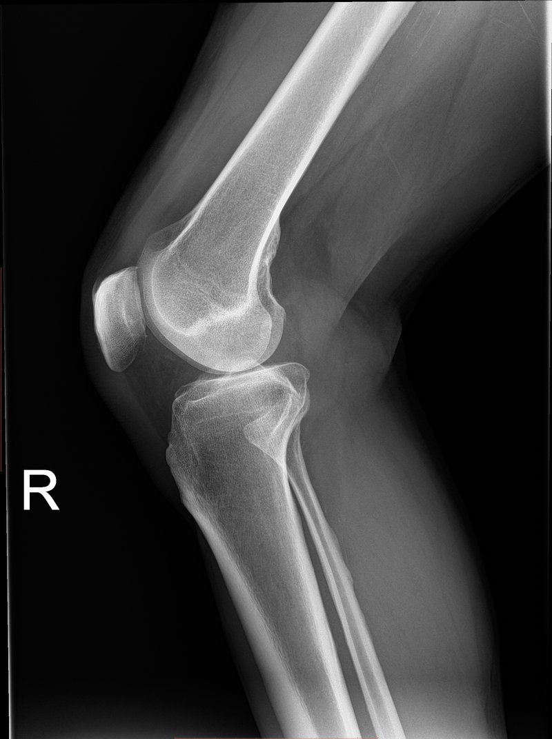
Position: Place the affected side nearest the IR and flex a routine, nontraumatic knee about 20–30°. Align the femoral epicondyles perpendicular to the IR. Flexion is primarily for a diagnostic lateral and is not a guarantee that every joint space will “open.”
Central ray: Angle 5–7° cephalad and center about 2.5 cm (1 inch) distal to the medial epicondyle. The cephalad angle compensates for the lower position of the medial femoral condyle in a mediolateral projection.
Criteria: Femoral condyles should be closely superimposed, the patella in profile, and the patellofemoral and tibiofemoral spaces visualized. The fibular head is partly superimposed by the tibia. Do not diagnose rotation from the visibility of the adductor tubercle alone; use condylar superimposition and the complete image.
AP Oblique Knee
Use about 45° rotation only when an oblique is ordered and the patient can tolerate it. Medial (internal) rotation best demonstrates the lateral femoral/tibial condyles and opens the proximal tibiofibular joint, with the fibular head relatively free of tibial superimposition. Lateral (external) rotation best demonstrates the medial condyles; the fibular head is more superimposed. Center about 1.3 cm distal to the patellar apex, normally with a perpendicular CR under the local protocol.
Special Projections: Names Are Not Interchangeable
Camp-Coventry Intercondylar View
This PA axial projection demonstrates the femoral intercondylar fossa, tibial intercondylar eminence, and posterior condylar surfaces. Place the patient prone and flex the knee 40–50° so the lower leg forms the same angle to the table. Direct the CR perpendicular to the lower leg—therefore approximately 40–50° caudad—to the popliteal crease/knee joint. The fossa should appear open and centered, without rotation.
Camp-Coventry = prone; it is not the supine IR-on-thigh method. Holmblad (kneeling) and Béclère (supine) are different intercondylar methods with different geometry. Never substitute one method's flexion or CR angle for another, and do not force any of them after acute trauma.
Merchant Bilateral Tangential Patellofemoral View
The Merchant method evaluates patellofemoral alignment and joint spaces. The patient is supine with both knees flexed 40° over the end of the table on a dedicated support; the IR is supported distal to the knees. Direct the CR 30° caudad from horizontal through both patellofemoral joints, according to the device and department protocol.
Live-verification marker: Merchant direction corrected
MERCHANT = 40° knee flexion + 30° CAUDAD beam—not 30° cephalad. “Sunrise,” “skyline,” and “tangential patella” are umbrella terms, not permission to mix Merchant, Settegast, Hughston, or inferosuperior positioning instructions.
Criteria: Both patellae and patellofemoral joint spaces should be shown without rotation; femoral condyles should be symmetric. Mark laterality unambiguously. A tangential patellar view requires substantial flexion and should not be attempted in suspected acute patellar fracture or dislocation unless specifically cleared and tolerated.
| Projection/method | Position | CR | Primary demonstration |
|---|---|---|---|
| AP, non-weight-bearing | Extended as tolerated | 1.3 cm distal to apex; habitus adjustment 3–5° | Distal femur, proximal tibia/fibula, tibiofemoral joint |
| Mediolateral | 20–30° flexion if nontraumatic | 5–7° cephalad; 2.5 cm distal to medial epicondyle | Patella in profile; superimposed femoral condyles |
| AP oblique | 45° medial or lateral rotation | Perpendicular; 1.3 cm distal to apex | Medial rotation opens proximal tibiofibular joint |
| Rosenberg PA weight-bearing | Bilateral, 45° flexion | 10° caudal | Weight-bearing tibiofemoral joint spaces |
| Camp-Coventry PA axial | Prone, 40–50° flexion | Perpendicular to lower leg (40–50° caudal) | Intercondylar fossa and eminence |
| Merchant bilateral tangential | Supine, 40° flexion | 30° caudal from horizontal | Patellofemoral alignment and joint spaces |
Technical Factors and Radiation Safety
- SID: Use the department's validated protocol; 100–110 cm (40–44 inches) is common for table/bucky knee work. Specialized standing or Merchant equipment may prescribe another SID.
- Exposure: Do not copy a universal kVp/mAs recipe. Select the current technique chart for the projection, patient thickness, detector, grid status, and SID. Use the lowest exposure that provides diagnostic quality and collimate tightly.
- Grid/AEC: An average adult knee is commonly imaged tabletop without a grid; use a grid only when part thickness/scatter and the departmental chart require it (often around 10–12 cm or more). AEC is generally unnecessary for a tightly collimated small joint unless a validated bucky protocol specifies it.
- Shielding: Do not place contact shielding in or near the field as routine practice. It can obscure anatomy, trigger repeats, or interfere with AEC. Follow current facility policy and explain that careful collimation and correct technique—not a shield outside the field—control dose.
- Identification: Confirm patient, side, indication, pregnancy status when relevant under local policy, and whether weight-bearing is requested and safe. Place the side marker before exposure.
Choosing the Next Modality
Radiography is usually the initial imaging test for chronic knee pain and is appropriate after acute trauma when validated clinical criteria indicate imaging. If radiographs are negative but occult fracture or internal derangement remains suspected, MRI without contrast is commonly the next study; CT without contrast is useful for detailed assessment of complex osseous fractures. Ultrasound may answer selected superficial tendon, bursal, cyst, or effusion questions but is not a replacement for radiographs or MRI for routine intra-articular assessment. The radiologist/ordering clinician and ACR criteria should guide escalation.
Quick Reference
- AP: epicondyles parallel → center 1.3 cm distal to patellar apex → use the protocol's habitus-based angle.
- Lateral: affected side down → 20–30° only if safe → 5–7° cephalad → center 2.5 cm distal to medial epicondyle.
- Camp-Coventry: prone → flex 40–50° → CR perpendicular to lower leg, to popliteal crease.
- Merchant: bilateral supine → flex 40° on support → CR 30° caudad from horizontal.
- Trauma: support as found; never force standing, flexion, rotation, kneeling, or tangential positioning.
Sources and Protocol Notes
Angles and centering vary among named methods and institutions. The positioning values above are conventional educational values; the Merchant description is corroborated by Idaho State University's radiographic-science article. Always use the named method and your institution's current protocol rather than blending techniques.
- Dartmouth Human Anatomy Learning Environment — Imaging the Knee and Leg (academic overview of standard views and MRI/CT roles)
- Idaho State University, Radiologic Technology — Tangential Projection of the Patella (Merchant: 40° flexion, 30° caudad; institutional educational source)
- Gundersen Health System — Knee Tunnel (Camp-Coventry) imaging protocol (institutional protocol: prone, 40° flexion, 40° caudal CR)
- ACR Appropriateness Criteria — Acute Trauma to the Knee
- ACR Appropriateness Criteria — Chronic Knee Pain
- CARES committee (AAPM/ACR/ASRT and partner organizations) — Patient Shielding FAQs
- ARRT — Position on Gonadal and Fetal Shielding (follow institutional policy; education and radiation protection remain required)