Paranasal Sinuses X-Ray Positioning: 5 Views (Waters, Caldwell, Lateral, SMV, Open-Mouth)
Dedicated paranasal sinus radiographs are now infrequently indicated. Uncomplicated acute rhinosinusitis is diagnosed clinically and ordinarily needs no imaging; when imaging is warranted for recurrent or chronic disease, surgical planning, suspected complications, invasive fungal disease, or a mass, CT or MRI is selected according to the clinical question. If a sinus radiographic series is specifically ordered under local protocol, its projections use a horizontal central ray, with the patient erect whenever safely possible, to preserve gravity-dependent air-fluid levels.
This guide explains five recognized sinus techniques — Waters (parietoacanthial), Caldwell (PA axial), lateral, SMV (submentovertical), and open-mouth Waters. They are not a universal five-view routine: many institutional protocols use three views (Waters, Caldwell, and lateral), while educational specifications also include horizontal-beam SMV. Always perform the projections on the order and in the department protocol.
If you are reviewing related facial and cranial imaging, pair this article with our Facial Bones X-Ray Positioning Guide and Skull X-Ray Positioning Guide, which share many of the same positioning lines and reference points. For patient preparation and communication strategies, see Patient Care in Radiography.
ARRT Memory Hook
Horizontal beam + erect patient (when safe). This combination demonstrates gravity-dependent air-fluid interfaces. An interface is a nonspecific finding: secretions and blood can both produce one, so it does not by itself prove infection, inflammation, or fracture. Never sit up or hyperextend a patient whose trauma or cervical-spine precautions prohibit it.
Paranasal Sinus Anatomy
The paranasal sinuses are air-filled cavities within the bones of the face and skull, lined by respiratory mucosa. They communicate with the nasal cavity through ostia and develop progressively from birth through adolescence. There are four sinus groups, generally bilateral but often asymmetric, each named for the bone that contains it:
| Sinus Group | Bone | Location | Best Demonstrated By |
|---|---|---|---|
| Frontal | Frontal bone | Lower forehead, above the orbits | Caldwell (PA axial) |
| Maxillary | Maxillae (paired) | Cheekbones, inferior to the orbits | Waters (parietoacanthial) |
| Ethmoid | Ethmoid bone | Multiple air cells between the orbits | Caldwell (anterior cells); SMV (posterior cells) |
| Sphenoid | Sphenoid bone | Behind the ethmoids, inferior to the sella turcica | Lateral; SMV; open-mouth Waters |
The frontal and maxillary sinuses and anterior/middle ethmoid cells drain toward the middle meatus; posterior ethmoid cells drain to the superior meatus, and the sphenoid sinus drains into the sphenoethmoidal recess. Ostial obstruction can impair ventilation and drainage, but radiographic mucosal opacity or a fluid level is not specific for bacterial infection.
Sinus development is age-dependent and variable: maxillary and ethmoid air cells are present at birth, whereas the sphenoid and especially frontal sinuses pneumatize later and mature through childhood or adolescence. Pediatric imaging must therefore be interpreted in an age-appropriate clinical context; when imaging is justified, modality selection follows the indication rather than age alone.
The Golden Rule: Upright + Horizontal Beam
For a dedicated conventional sinus series intended to assess fluid levels, the explanation is straightforward:
- Erect positioning allows fluid or blood to layer dependently, potentially creating a visible air-fluid interface. In a supine projection, dependent posterior layering may appear as opacity rather than a horizontal interface.
- Horizontal central ray preserves that horizontal interface. In the sinus Caldwell, create the required 15° OML-to-ray relationship by positioning the head/receptor—not by angling the beam away from horizontal.
If a patient cannot stand, seated erect positioning may be possible. If the patient must remain recumbent, use only a department-approved horizontal-beam adaptation; do not remove immobilization or manipulate the neck merely to obtain a sinus projection. Suspected facial fractures and acute trauma are generally evaluated with maxillofacial CT rather than a routine sinus series.
📝 Clinical Pearl
Safety overrides textbook geometry. Keep the beam horizontal in an approved recumbent adaptation, but do not claim that one substitute view is universally diagnostic. Confirm the order/protocol, maintain spinal precautions, and escalate to CT when trauma or the required anatomy cannot be adequately assessed radiographically.
Positioning Lines Reference
Four positioning references are used repeatedly in sinus radiography and appear in radiography education:
| Line | Abbreviation | Definition | Used In |
|---|---|---|---|
| Orbitomeatal line | OML | Outer canthus (lateral eye) to EAM | Caldwell, Waters, lateral |
| Mentomeatal line | MML | Mentum (chin) to EAM | Waters (perpendicular to IR) |
| Infraorbitomeatal line | IOML | Infraorbital margin to EAM | SMV, lateral |
| Midsagittal plane | MSP | Divides skull into symmetric left and right halves | All views (rotation check) |
Projection 1: Waters (Parietoacanthial) — Maxillary Sinuses
The Waters projection is the conventional radiographic view that best demonstrates the maxillary sinuses. It projects the petrous ridges just below the maxillary sinus floors, reducing superimposition over the maxillary antra.
| Parameter | Waters (Parietoacanthial) Setup |
|---|---|
| Patient position | Upright, facing the upright detector. The chin is placed against the IR with the neck extended. |
| Key alignment | MSP perpendicular to IR (no rotation or tilt). Extend the neck until the MML (mentomeatal line) is perpendicular to the IR; the OML is then approximately 37° to the plane of the IR. |
| Central ray | Horizontal (0° angle), exiting at the acanthion (junction of the upper lip and nasal septum). |
| SID | 40 inches (100 cm). |
| Collimation | Include the frontal sinuses superiorly, maxillary sinuses inferiorly, and both orbits laterally. |
| Respiration | Suspended expiration. |
Waters Evaluation Criteria
- Petrous ridges are projected immediately below the maxillary sinus floors — if they appear higher, the chin was not extended enough; if much lower, the chin was over-extended.
- Orbits and maxillary sinuses appear symmetric side to side (no rotation).
- The nasal septum is aligned at the midline.
- Frontal sinuses are visible above the frontonasal suture.
- The image demonstrates air-fluid levels if present, with sharp sinus wall margins.
Open-Mouth Waters (Sphenoid Sinuses)
The open-mouth Waters uses the same head and CR position as the standard Waters, but the patient opens widely without changing head extension. This projects the sphenoid sinus through the open oral cavity. It requires less extension than an SMV, but no projection requiring neck extension should be attempted when cervical motion is contraindicated.
- CR: Same as Waters — horizontal, exiting at the acanthion.
- Evaluation: Sphenoid sinuses are visible through the open mouth. Petrous ridges remain below the maxillary floors. The maxillary sinuses are also well demonstrated.
- Common error: If the patient closes the mouth during exposure or does not open widely enough, the sphenoid sinuses will be obscured by the mandible.
Projection 2: Caldwell (PA Axial) — Frontal and Ethmoid Sinuses
The Caldwell view demonstrates the frontal sinuses and anterior ethmoid air cells. For a sinus study, the beam stays horizontal; a 15° relationship between the OML and horizontal CR projects the petrous ridges into the lower third of the orbits.
| Parameter | Caldwell (PA Axial) Setup |
|---|---|
| Patient position | Erect, facing a vertical detector, with the neck adjusted so the OML is 15° above the horizontal ray. A department may instead use a correctly aligned 15° receptor/grid setup while keeping the beam horizontal. |
| Key alignment | MSP perpendicular to IR, with no rotation or tilt. OML 15° to the horizontal CR (or perpendicular to a protocol-approved tilted receptor). |
| Central ray | Horizontal, exiting at the nasion. Do not use the 15° caudad tube angle of a routine skull Caldwell when the purpose is to preserve sinus fluid levels. |
| SID | 40 inches (100 cm). |
| Respiration | Suspended expiration. |
Caldwell Evaluation Criteria
- Petrous ridges are projected in the lower third of the orbits.
- Frontal sinuses are visible above the frontonasal suture.
- The orbits and innominate lines appear symmetric (no rotation).
- A horizontal line through the petrous ridges should be level (no tilt).
- Anterior ethmoid air cells are visible inferior to the frontal sinuses.
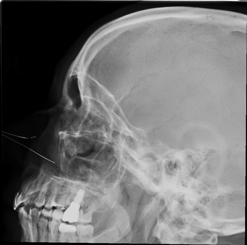
Projection 3: Lateral — All Four Sinus Groups
The lateral sinus view includes all four sinus groups on one image and demonstrates the sphenoid sinus and sella turcica in profile. A routine sinus protocol generally obtains one lateral because right and left sinuses are superimposed; obtain another side only when the order or local protocol specifically requires it.
| Parameter | Lateral Sinus Setup |
|---|---|
| Patient position | Upright, seated or standing. The side of interest is placed closest to the detector. The body may be rotated slightly for comfort, but the head must be in a true lateral position. |
| Key alignment | MSP parallel to IR. Interpupillary line (IPL) perpendicular to IR. IOML parallel to the transverse axis of the IR (i.e., the IOML is horizontal). |
| Central ray | Horizontal and perpendicular to the IR, centered to the zygoma midway between the outer canthus and EAM (approximately 2 cm posterior to the outer canthus in an average adult). |
| SID | Use the department protocol (commonly 40 inches/100 cm). |
| Respiration | Suspended respiration. |
Lateral Evaluation Criteria
- Sella turcica is seen in true profile — no double contours.
- Orbital roofs and mandibular rami are superimposed (no tilt or rotation).
- All four sinus groups are visible: frontal anteriorly, maxillary inferiorly, ethmoid medially, and sphenoid posteriorly.
- The sphenoid sinus is optimally demonstrated.
- Sharp bony outlines with no motion blur.
Projection 4: SMV (Submentovertical) — Sphenoid and Ethmoid
The full-basal SMV provides an axial view of the skull base and demonstrates the sphenoid sinus and posterior ethmoid air cells. It requires marked neck extension and must not be attempted when cervical-spine injury or unsafe motion is suspected.
| Parameter | SMV Setup |
|---|---|
| Patient position | Erect with the vertex against the vertical IR. Extend the neck only if safe until the IOML is parallel to the IR. A supine vertical-beam skull SMV does not preserve air-fluid levels and is not equivalent to this sinus projection. |
| Key alignment | MSP perpendicular to IR. IOML parallel to IR. No rotation or tilt. |
| Central ray | Horizontal, perpendicular to the IOML, entering on the MSP approximately 3/4 inch (2 cm) anterior to the level of the EAM and directed toward the vertex. |
| SID | 40 inches (100 cm). |
| Respiration | Suspended respiration, mouth closed. |
SMV Evaluation Criteria
- Mandibular condyles are projected anterior to the petrous ridges.
- The mentum is projected anterior to the ethmoid sinuses.
- Equal distance from the mandible to the lateral skull borders (no rotation).
- Sphenoid sinus and posterior ethmoid air cells are visualized.
- The mandibular symphysis does not obscure the ethmoid region (indicates adequate extension).
⚠️ Common SMV Pitfall
Inadequate neck extension places the mandible over the region of interest. If extension is merely limited but safe, an open-mouth Waters may demonstrate the sphenoid sinus with less extension. If motion is contraindicated or anatomy remains obscured, stop rather than force the position and follow the protocol for alternative imaging.
Exposure, Grid, and Radiation Safety
- Technique: use the current department technique chart for the detector, generator, patient size, and projection. A fixed kVp/mAs recipe is not transferable between screen-film, computed radiography, and digital radiography systems. Use the shortest practical exposure time and assess exposure-index feedback to avoid dose creep.
- Grid: grid use is protocol- and equipment-dependent. If a grid is used—particularly with a tilted receptor—center and align the tube to that grid to prevent cutoff. Do not state that sinus imaging always or never requires a grid.
- Collimation: collimate to the required sinus anatomy and avoid unnecessary direct irradiation of the ocular lenses. Remove glasses, removable dental appliances, earrings, hairpins, and other artifacts when they overlap the field.
- Patient shielding: routine gonadal or fetal contact shielding is no longer recommended by the AAPM because it provides negligible benefit and can obscure anatomy, interfere with automatic exposure control, or prompt repeats. Follow current law and facility policy; never place shielding in the primary field.
- Pregnancy: ask and document pregnancy status according to facility policy. Because the pelvis is outside a properly collimated sinus beam, conceptus exposure is limited to scatter; pregnancy alone is not a reason to withhold a medically necessary head examination. Confirm justification and optimize the exam rather than automatically canceling or adding abdominal shielding.
Current Clinical Role
Plain sinus radiography is not a screening test for routine uncomplicated sinus symptoms. ACR guidance rates paranasal sinus radiography “usually not appropriate” for the evaluated sinonasal-disease scenarios. No imaging is usually needed for uncomplicated acute rhinosinusitis; noncontrast maxillofacial CT is generally appropriate for recurrent/chronic disease or surgical planning, while suspected orbital/intracranial complication, invasive fungal disease, or tumor may require contrast-enhanced CT and/or MRI. Use the examination selected by the radiologist/referrer for the actual clinical scenario.
Quick Reference Table
| Projection | Best For | CR Entry/Exit | Key Positioning Check |
|---|---|---|---|
| Waters | Maxillary sinuses | Horizontal, exits acanthion; MML ⟂ IR | Petrous ridges just below maxillary floors |
| Open-mouth Waters | Sphenoid sinuses (alternative to SMV) | Horizontal, exits acanthion; patient opens mouth | Sphenoids through open mouth |
| Caldwell (PA axial) | Frontal + anterior ethmoid | Horizontal, exits nasion; OML 15° to CR | Petrous ridges in lower 1/3 of orbits |
| Lateral | All four groups; sphenoid in profile | Horizontal, zygoma midway from outer canthus to EAM | Sella turcica in profile; rami superimposed |
| SMV | Sphenoid + posterior ethmoid; skull base | Horizontal, ⟂ IOML, enters 3/4″ (2 cm) anterior to EAM | Condyles anterior to petrous ridges |
Common Positioning Errors
| Error | How It Looks | Correction |
|---|---|---|
| Waters: petrous ridges too high | Petrous ridges overlap the maxillary sinus floors | Extend chin further, if safe, until MML is ⟂ to IR |
| Waters: petrous ridges too low | Petrous ridges far below maxillary floors; sinuses foreshortened | Chin over-extended — reduce extension slightly |
| Caldwell: petrous ridges too high | Petrous ridges fill mid-to-upper orbits, obscuring ethmoid region | Increase the OML-to-horizontal-CR relationship toward 15° by adjusting head/receptor—not tube angle |
| Lateral: orbital roofs not superimposed | Double contour of orbital roofs | Check for tilt (IPL not ⟂ to IR) |
| SMV: mandible obscures ethmoids | Mandible overlaps the ethmoid air cells | If neck motion is safe, increase extension until IOML is ∥ to IR; otherwise stop |
| Rotation | Asymmetry or unwanted double contours | Check MSP: perpendicular for PA/Waters/SMV, parallel for lateral |
| Angled CR (sinus series) | A fluid interface may be obscured or distorted | Keep CR horizontal; obtain Caldwell geometry with head/receptor positioning |
Registry Review Pearls
- ARRT's published Radiography Content Specifications list horizontal-beam lateral, Caldwell, Waters, and full-basal SMV under paranasal sinuses. This is an examination content outline, not a mandate that every clinical department perform a four-view routine.
- Dedicated sinus projections use a horizontal central ray to preserve air-fluid interfaces.
- The Waters view best demonstrates the maxillary sinuses radiographically — the petrous ridges should be just below the maxillary floors.
- The Caldwell view evaluates frontal and anterior ethmoid sinuses — the petrous ridges must be in the lower third of the orbits.
- The lateral view best shows the sphenoid sinus and sella turcica in profile.
- The SMV requires the IOML to be parallel to the IR; inadequate extension causes mandibular superimposition.
- The MML (mentomeatal line), not the AML, is perpendicular to the IR for Waters; this places the OML about 37° to the IR.
- Perform sinus studies erect when safely possible; maintain trauma and cervical-spine precautions.
- Plain radiography has a limited modern role and should not be described as routine screening. ACR criteria favor no imaging for uncomplicated acute rhinosinusitis and CT/MRI for indicated detailed evaluation.
- Protect the radiosensitive ocular lenses primarily through justification, protocol optimization, accurate positioning, and tight collimation—not by placing a shield over required anatomy.
📖 Evidence and Protocol Note
No proprietary textbook attribution is asserted. Geometry and evaluation criteria were cross-checked against ARRT's published positioning outline and accredited/institutional image standards; current clinical-use statements follow ACR sinonasal guidance. Local protocols govern the ordered views, exposure factors, grid, and safe alternatives. For a broader critique framework, see our Image Critique and Evaluation Methodology guide.
- ACR Appropriateness Criteria®: Sinonasal Disease
- ARRT Radiography Examination Content Specifications (paranasal sinus projections)
- Dartmouth Geisel School of Medicine: Digital Image Standards for Routine Radiography
- Medford Radiological Group: Paranasal Sinuses X-ray Guideline (three-view institutional example)
- AAPM Position Statement on Patient Gonadal and Fetal Shielding
- ACR–SPR Practice Parameter for Imaging Pregnant or Potentially Pregnant Patients
Test Your Knowledge
Click an option to check your answer. Correct answers turn green; incorrect answers turn red and still reveal the explanation.
ARRT's positioning outline specifies a horizontal beam for the listed sinus projections so a gravity-dependent air-fluid interface is preserved. For example, the sinus Caldwell keeps the beam horizontal and creates the 15° OML relationship through head/receptor positioning; a routine skull Caldwell commonly angles the CR.
A correctly positioned Waters projection places the petrous ridges immediately below the maxillary sinus floors. If the ridges overlap the maxillary sinuses, the chin was not extended enough. Option C describes the Caldwell view, not the Waters. Option A describes the lateral view, and option B describes the SMV.
The open-mouth Waters maintains the standard Waters head and horizontal-CR position while the patient opens widely, projecting the sphenoid sinus through the oral cavity. It requires less extension than the SMV. Do not extend the neck at all if cervical motion is contraindicated; use the approved alternative imaging pathway instead.
With the sinus Caldwell's horizontal beam and 15° OML-to-CR relationship, the petrous ridges project into the lower third of the orbits. Do not substitute the caudally angled CR used for a routine skull Caldwell when fluid levels are the target. Option A describes the SMV; B and D are lateral criteria.
The lateral sinus view best demonstrates the sphenoid sinus, along with the sella turcica in profile. It also shows all four sinus groups on a single image. The Waters is best for maxillary sinuses, and the Caldwell is best for frontal and anterior ethmoid sinuses.