Scatter Radiation from Portable X-Ray: Does It Affect the Room — and Do the Benefits Outweigh It?
The short answer
Yes, portable X-ray produces scatter radiation that reaches the people surrounding the patient — but no, it does not need to be avoided for that reason.
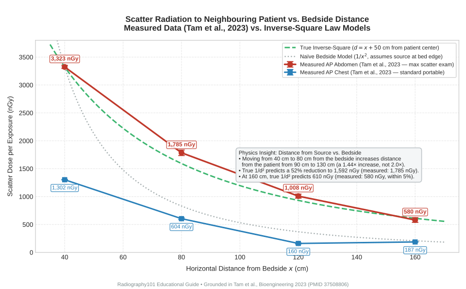
Measured at clinically realistic geometry, the scatter reaching a neighbouring patient from an adult AP abdomen exposure (the highest-scatter bedside exam) falls from 3 323 nGy per exposure at 40 cm from the bedside to 580 nGy at 160 cm — an 83 % reduction (Tam et al., 2023). For a routine portable chest radiograph, the scatter dose is even lower: 1 302 nGy at 40 cm, dropping to just 187 nGy at 160 cm (Tam et al., 2023). In a neonatal intensive care unit (NICU), the cumulative scatter dose to an adjacent patient or their caregiver was estimated at no more than 40 µSv over an entire three-month admission, while annual staff scatter dose averaged 160 µSv per year — compared to the occupational dose limit of 20 mSv per year (Longo et al., 2018).
Meanwhile, the reason we perform imaging at the bedside rather than sending the patient to the radiology department is that patient transport itself carries substantial, measurable clinical risk. In a multicentre cohort of 3 006 intrahospital transports of ventilated ICU patients, post-transport complications occurred in 37.4 % of patients (Schwebel et al., 2013). In another prospective series, adverse events occurred during 45.8 % of transports, with 16.8 % classified as serious (Parmentier-Decrucq et al., 2013).
The evidence-based position is not "portable imaging is dangerous; avoid it." Rather:
The evidence-based position
Portable radiography is justified when a patient cannot be transported safely to the radiology department — that is the primary clinical benefit. The scatter radiation it creates in the surrounding room is minimal, orders of magnitude below regulatory limits at standard working distances, and easily controlled through collimation, distance, and shielding. Distance is your most effective tool, and the minor scatter risk is readily managed for everyone in the room.
Below is the clinical dosimetry evidence, a corrected distance calculation addressing a classic physics trap, and a practical pre-exposure bedside checklist.
Why portable imaging scatters differently from a fixed room
A fixed radiographic room is engineered around the principle of containment: lead-lined walls, a dedicated Bucky assembly, and a shielded control booth for staff.
Bedside portable imaging alters these conditions:
- Lack of permanent structural shielding: Imaging takes place in an open bay, intensive care unit, or trauma room where adjacent patients cannot always be relocated.
- Horizontal and multi-directional beam geometry: While portable chest exams are generally performed anteroposterior (AP) with the patient supine or semi-erect, decubitus and cross-table projections direct the beam and resulting scatter horizontally across the room rather than down into a lead-lined floor or Bucky tray.
- Shorter source-to-image distance (SID): Portable exams often use a 100 cm SID compared to the standard 180 cm SID used for dedicated upright chest radiography. To achieve adequate detector exposure at a shorter SID, entrance skin exposure is adjusted, and the patient's body becomes the primary three-dimensional source of scattered Compton photons.
- Field size and collimation challenges: In critical care environments crowded with lines, monitors, and ventilators, technologists may inadvertently open the collimator wider than necessary. Because scatter production is directly proportional to irradiated tissue volume, poor collimation significantly increases the scatter field in the room.
These factors do not mean portable exams are unsafe; rather, they highlight why technique, collimation, and distance dictate whether bystander dose remains negligible.
What clinical measurements demonstrate
Scatter dosimetry is reported across a variety of units in the literature (nGy, µGy, µSv, mSv). For diagnostic X-rays, the radiation weighting factor (wR) is 1.0, meaning 1 µGy of absorbed dose equals 1 µSv of equivalent dose.
The table below compiles measured scatter values across peer-reviewed studies:
| Scenario / measurement | Exposure parameters / context | Measured scatter dose | Source |
|---|---|---|---|
| Nearby patient, 40 cm from bedside | AP Abdomen (81 kVp, 11 mAs, adult phantom) | 3 323 ± 28 nGy per exposure | Tam et al., 2023 (PMID 37508806) |
| Nearby patient, 80 cm from bedside | AP Abdomen (81 kVp, 11 mAs, adult phantom) | 1 785 ± 50 nGy per exposure | Tam et al., 2023 |
| Nearby patient, 160 cm from bedside | AP Abdomen (81 kVp, 11 mAs, adult phantom) | 580 ± 42 nGy per exposure | Tam et al., 2023 |
| Nearby patient, 40 cm from bedside | AP Chest (81 kVp, 11 mAs, adult phantom) | 1 302 ± 4 nGy per exposure | Tam et al., 2023 |
| Nearby patient, 80 cm from bedside | AP Chest (81 kVp, 11 mAs, adult phantom) | 604 ± 14 nGy per exposure | Tam et al., 2023 |
| Nearby patient, 160 cm from bedside | AP Chest (81 kVp, 11 mAs, adult phantom) | 187 ± 6 nGy per exposure | Tam et al., 2023 |
| Caretaker / parent, 140 cm standing height | AP Chest/Abdomen (65 kVp, 3.2 mAs, pediatric) | 659 ± 7 nGy per exposure | Tam et al., 2023 |
| Caretaker / parent, 50 cm seated/crouched | AP Chest/Abdomen (65 kVp, 3.2 mAs, pediatric) | 33 ± 1 nGy per exposure | Tam et al., 2023 |
| Technologist, > 150 cm from tube/patient | Mobile chest radiography | < 1 µSv per exposure | Otomo et al., 2023 (PMID 36829753) |
| NICU staff dose, per examination | Bedside neonatal radiography | 0.012 – 0.095 µSv per exam | Longo et al., 2018 (PMID 29442153) |
| NICU annual staff scatter dose | Typical NICU annual workload | ≈ 160 µSv / year (0.16 mSv/yr) | Longo et al., 2018 |
| NICU adjacent infant / caregiver | Full 3-month hospitalization stay | ≤ 40 µSv total (0.04 mSv) | Longo et al., 2018 |
| Nursery crib spacing, at 1 m | Neonatal lateral skull (maximum scatter) | ≈ 0.05 µGy per exposure | Burrage et al., 2003 (PMID 12861430) |
| NICU adjacent isolette, 93 cm & 125 cm | Neonatal chest at 56 kVp (2–3 ft spacing) | Minimal compared to 8.493 µGy daily background | Trinh et al., 2010 (PMID 19997727) |
| Neonatal incubator lead shield | 0.5 mm Pb shield at 60–90 kVp | Scatter reduced by 84 – 92 % at 1 m | Cheewakul et al., 2026 (PMID 42144420) |
| Technologist eye lens dose | Mobile radiography workload (neck badge) | Up to 0.3 mSv / month (lead glasses cut ~45 %) | Matsumoto et al., 2023 (PMID 36418072) |
| CR to Flat-Panel Detector (FPD) | Mobile radiography transition | Technologist lens dose reduced by 66 % | Radiol Phys Technol, 2025 (PMID 40553226) |
Figure 1 illustrates the measured scatter values across distances alongside the theoretical inverse-square predictions.
Note on the 80 cm value: the paper’s abstract reports 1 785 nGy while its Table 4 reports 1 788 ± 50 nGy for the same AP abdomen point. This article and the figure use the abstract value (1 785 nGy) consistently; the difference (0.2 %) does not affect any conclusion.
Key clinical insights from the data
- Patient dose dwarfs neighbour dose: While entrance skin dose to the patient being imaged is roughly 15 to 74 µGy for a neonatal exam (Yu, 2010, PMID 21146794) and several hundred µGy to a few mGy for an adult exam, the neighbour receives less than 1 % of that dose per exposure — for an adult AP abdomen, about 3.3 µGy of scatter 40 cm from the bedside against a patient entrance skin dose of several hundred µGy; for neonates, of the order of 0.05 µGy per exposure at 1 m (Burrage et al., 2003) against a 15–74 µGy entrance skin dose.
- Technologists accumulate occupational dose over careers: A single exposure delivers negligible scatter (< 1 µSv) to an operator standing at 1.5–2 meters, but performing hundreds of mobile exams annually accumulates occupational dose. Advances such as the transition from computed radiography to digital flat-panel detectors reduced technologist lens dose by 66 % (PMID 40553226), and lead glasses provide an additional 45 % lens dose reduction (Matsumoto et al., 2023).
- Scatter fields are three-dimensional: In pediatric exams, caretaker dose dropped from 659 nGy at eye level (140 cm) to 33 nGy at low level (50 cm) — a 20-fold difference — scatter dose varies strongly with height as well as distance. Staying low or positioning behind barriers provides substantial protection.
Regulatory dose limits in perspective
International radiation protection standards set clear annual limits:
- Public Dose Limit: 1 mSv (1 000 µSv) per year (IAEA GSR Part 3, Schedule III.3).
- Occupational Dose Limit: 20 mSv (20 000 µSv) per year, averaged over 5 consecutive years, with no single year exceeding 50 mSv (IAEA GSR Part 3, Schedule III.1).
Comparing the measured bedside scatter data against these thresholds:
- An adjacent infant or visitor in a NICU accumulating ≤ 40 µSv over 3 months receives only 4 % of the annual public limit (and less than a week's worth of typical natural background radiation).
- A staff member receiving 160 µSv per year from mobile scatter uses only 0.8 % of the occupational limit.
Crucial distinction
Crucial Distinction: As stated in ICRP Publication 105, dose limits do not apply to medical exposures of the patient being imaged. Patient care is governed strictly by justification (does the exam provide more medical benefit than harm?) and optimisation (is the dose as low as reasonably achievable, ALARA, for the required image quality?).
The distance question: bedside geometry vs. the inverse-square law
A frequent source of confusion on radiography exams and in clinical radiation safety calculations is how the inverse-square law applies to bedside scatter.
The bedside distance trap
The inverse-square law (I2 = I1 × (d1 / d2)2) requires that distances d1 and d2 be measured from the radiation source (the patient's irradiated body), not from the edge of the bed.
In the phantom study by Tam et al. (2023), the measurement coordinate x represented the distance from the bedside, while the bed half-width from the midline of the patient to the edge of the mattress was 50 cm.
- At x = 40 cm from the bedside, true distance from the source is d1 = 40 + 50 = 90 cm.
- At x = 80 cm from the bedside, true distance from the source is d2 = 80 + 50 = 130 cm.
- At x = 160 cm from the bedside, true distance from the source is d3 = 160 + 50 = 210 cm.
Step-by-step calculation
Step 1: Check the distance ratio Moving from 40 cm to 80 cm from the bedside does not double the distance from the patient:
Step 2: Calculate theoretical inverse-square prediction at 80 cm
The actual measured dose was 1 785 nGy (a 46.3 % drop to 53.7 % of initial dose). The slight difference (1 785 vs 1 592 nGy) is attributable to extended scattering volume and ambient room scatter.
Step 3: Calculate theoretical inverse-square prediction at 160 cm
The actual measured dose was 580 ± 42 nGy — matching the theoretical inverse-square prediction within 5 %!
How many exposures to reach the 1 mSv public limit?
Using the measured AP abdomen scatter (conservative upper bound):
* At 40 cm bedside (3 323 nGy / 3.323 µGy):
* At 160 cm bedside (580 nGy / 0.58 µGy):
For routine portable chest exams (187 nGy at 160 cm), it would require more than 5 300 exposures to reach 1 mSv.
Evaluating the risk-benefit equation: transport risk vs. scatter dose
To determine whether portable radiography is justified, the minor scatter dose must be weighed against the alternative: transporting an unstable patient through hospital hallways and elevators to the central radiology department.
The clinical hazards of intrahospital transport
Intrahospital transport of critically ill patients involves disconnecting and reconnecting monitoring lines, transferring ventilators, and navigating corridors:
| Study | Cohort / sample | Documented adverse events & complications |
|---|---|---|
| Schwebel et al., 2013 (PMID 23863225) | 3 006 transports across 1 782 ventilated ICU patients | Post-transport complications occurred in 37.4 % of patients (pneumothorax, atelectasis, VAP, glycemic instability; OR = 1.9). |
| Parmentier-Decrucq et al., 2013 (PMID 23587445) | 262 transports across 184 ICU patients | Adverse events in 45.8 % of transports; 26.0 % directly impacted the patient; 16.8 % were serious clinical events. |
| Critical Care Review, 1999 (PMID 11094486) | Systematic analysis | Adverse effects in up to 70 % of transports; 12 % experienced sustained respiratory deterioration. |
| Austrian Cohort, 2007 (PMID 17671822) | 452 transports across 226 patients | Critical incident rate of 4.2 % under specialized transport team protocols. |
While event definitions vary (ranging from monitor disconnects to hemodynamic collapse), moving a critically ill patient is a major clinical intervention. Conversely, transports performed for diagnostic procedures changed patient management in 40 % to 50 % of cases (Waydhas, 1999, PMID 11094486), and bedside chest examinations are judged clinically useful in 76 % to 94 % of intensive-care cases (Wandtke, 1994, PMID 8043058).
Weighing the trade-off
- For the patient: Bedside imaging eliminates the high risk of intrahospital transport complications while providing critical diagnostic guidance.
- For room occupants and neighbouring patients: Scatter doses of 0.2 to 3 µGy per exam represent no more than 0.3 % of the annual public dose limit. With basic distance (standing back 2 meters) and shielding, exposure is reduced to near-background levels.
Conclusion
The clinical benefits of portable radiography substantially outweigh the minimal scatter radiation, provided the exam is clinically indicated and ALARA practices are followed.
Bedside Radiation Safety Checklist
Use this practical workflow whenever performing portable radiography in an occupied room or intensive care unit:
Pre-Exposure Planning
- Verify Clinical Justification: Confirm the specific clinical question and ensure a portable examination is indicated over department imaging.
- Select Optimized Factors: Match kVp and mAs to patient habitus; consider whether a physical grid is necessary or if digital processing / virtual grids can be utilized to reduce dose.
- Survey the Room: Identify all occupants, including neighbouring patients, visitors, nurses, and allied health staff.
Beam and Scatter Control
- Collimate Strictly: Collimate tightly to the anatomical area of interest. Reducing field size is the single most effective way to decrease scatter production.
- Direct the Beam Safely: Avoid directing the primary beam or central scatter axis toward adjacent occupied beds whenever feasible.
- Ensure Primary Beam Clearance: Verify that no bystanders, staff hands, or non-essential equipment are in the path of the primary beam.
Protecting Bystanders and Visitors
- Request Non-Essential Staff Step Out: Politely ask visitors and non-essential personnel to step outside the room during the exposure.
- Maximize Distance for Those Who Cannot Leave: Position neighbouring patients and caregivers as far from the tube and the patient as the room allows. Many programs use approximately 2 m (6 ft) as a practical reference when no barrier is available, but the definitive position follows your facility’s radiation-safety rules and any room survey.
- Utilize Physical and Height Shielding: Position lead mobile shields or incubator shields where available. Remind remaining helpers that staying low or seated behind bed frames reduces exposure.
Technologist Self-Protection
- Maximize Operating Distance: Extend the exposure switch cord fully and step back to the greatest practical distance, or stand behind the portable machine chassis. Where no barrier is available, roughly 2 m (6 ft) is a common program reference — follow your facility’s rule.
- Wear Prescribed PPE: Wear lead aprons (0.5 mm Pb equivalent) and thyroid shields in accordance with departmental policy, particularly when assisting with positioning close to the patient.
- Wear Dosimetry Badges Correctly: Wear each dosimeter exactly where your radiation safety officer or badge report specifies — placement differs between a single-badge and a double-badge program, so a universal “collar outside the apron” rule does not apply.
- Avoid Manual Restraint: Never hold cassettes or patients during exposure; use positioning sponges, detector holders, or mechanical aids.
Post-Exposure Review
- Check Image Quality Before Leaving: Review diagnostic acceptability immediately to avoid unnecessary repeat exposures.
- Document Exceptions: Record any unusual positioning constraints or shielding limitations in departmental logs.
Common Misconceptions vs. Clinical Evidence
| Common misconception | Evidence-based reality |
|---|---|
| "Scatter from mobile X-ray is negligible, so precautions are unnecessary." | While doses per exposure are small (< 3 µGy), radiation is real and cumulative. Distance alone cut measured scatter by about 83 % between 40 cm and 160 cm from the bedside in Tam et al. (2023), and collimation reduces the irradiated volume that produces scatter — both are free. |
| "The inverse-square law doesn't work for scatter because it's not a point source." | When distance is correctly measured from the scattering source (patient center) rather than the bedside edge, the inverse-square law predicts measured scatter within 5 % to 12 %. |
| "Standing in the corner of the room guarantees safety." | Distance and shielding provide protection; location in a room does not. If a corner is closer to the patient or unshielded, it offers less protection than standing 2 meters back behind the machine. |
| "The adjacent patient receives more radiation than the patient being imaged." | The imaged patient receives substantially more dose than anyone else in the room: in adults, several hundred µGy of entrance skin dose against about 3.3 µGy of scatter 40 cm from the bedside (a ratio of the order of 100:1); in neonates, a 15–74 µGy entrance skin dose against measured neighbour doses of the order of 0.05 µGy per exposure at 1 m (Burrage et al., 2003). |
| "Lead aprons are legally required for all room occupants during mobile X-rays." | Regulatory standards emphasize distance and shielding. Non-essential persons should step out; when distance and shielding are used, scatter exposure remains far below regulatory concern. |
Authoritative Sources
- Tam SY, Fung YY, Lau SY, Lam WN, Wong ET. Scatter Radiation Distribution to Radiographers, Nearby Patients and Caretakers during Portable and Pediatric Radiography Examinations. Bioengineering (Basel). 2023;10(7):779. PMID: 37508806.
- Longo F, Genovese E, Donatiello S, Cassano B, et al. Quantification of scatter radiation from radiographic procedures in a neonatal intensive care unit. Pediatr Radiol. 2018;48(5):715–721. PMID: 29442153.
- Burrage JW, Rampant PL, Beeson BP. Scatter and transmission doses from several pediatric X-ray examinations in a nursery. Pediatr Radiol. 2003;33(10):704–708. PMID: 12861430.
- Trinh AM, Schoenfeld AH, Levin TL. Scatter radiation from chest radiographs: is there a risk to infants in a typical NICU? Pediatr Radiol. 2010;40(5):704–707. PMID: 19997727.
- Otomo K, Inaba Y, Abe K, Onodera M, et al. Spatial Scattering Radiation to the Radiological Technologist during Medical Mobile Radiography. Bioengineering (Basel). 2023;10(2):259. PMID: 36829753.
- Matsumoto M, Takei Y, Matsuda T, Masuda T. Measurement of the Lens Dose for Radiological Technologists during Mobile Radiography. Nihon Hoshasen Gijutsu Gakkai Zasshi. 2023;79(1):38–45. PMID: 36418072.
- Chida K, et al. Assessment of lens absorbed dose by radiological technologists during mobile X-ray radiography: a comparison between computed radiography and flat panel detector systems. Radiol Phys Technol. 2025;18(3):766–774. PMID: 40553226.
- Cheewakul J, Ina N, Aonprawat S, Marshall SK, Chaichulee S. Radiation shield for neonatal incubators: design and dosimetric evaluation in NICU radiography. Sci Rep. 2026;16(1):11420. PMID: 42144420.
- Yu CC. Radiation safety in the neonatal intensive care unit: too little or too much concern? Pediatr Neonatol. 2010;51(6):311–319. PMID: 21146794.
- Schwebel C, Clec'h C, Magne S, Minet C, et al. Safety of intrahospital transport in ventilated critically ill patients: a multicenter cohort study. Crit Care Med. 2013;41(8):1919–1928. PMID: 23863225.
- Parmentier-Decrucq E, Poissy J, Favory R, Nseir S, et al. Adverse events during intrahospital transport of critically ill patients: incidence and risk factors. Ann Intensive Care. 2013;3(1):10. PMID: 23587445.
- Waydhas C. Intrahospital transport of critically ill patients. Crit Care. 1999;3(5):R83–R89. PMID: 11094486.
- Zuchegne E, et al. Incidence of complications in intrahospital transport of critically ill patients — experience in an Austrian university hospital. Wien Klin Wochenschr. 2007;119(17-18):556–561. PMID: 17671822.
- Wandtke JC. Bedside chest radiography. Radiology. 1994;190(1):1–10. PMID: 8043058.
- Bwanga O. What nurses need to know about mobile radiography. Br J Nurs. 2020;29(18):1064–1067. PMID: 33035087.
- International Commission on Radiological Protection (ICRP). Radiological Protection in Medicine. ICRP Publication 105. Ann ICRP. 2007;37(6):1–63.
- International Atomic Energy Agency (IAEA). Radiation Protection and Safety of Radiation Sources: International Basic Safety Standards. General Safety Requirements Part 3 (GSR Part 3). Vienna: IAEA; 2014.

Registered Radiologic Technologist (RRT)
Educational resource for radiologic technology students and professionals. This guide is for educational purposes and does not replace the imaging order, clinical assessment, institutional protocol, or the judgment of the responsible physician.
Check Your Understanding
Test your knowledge of mobile radiography scatter and clinical safety:
Answers and Detailed Explanations 1 — B. Distance is the most effective and immediately accessible protection tool. Why not A: Increasing mAs directly increases radiation output and scatter production. Why not C: Turning away does not significantly alter absorbed whole-body dose. Why not D: The technologist's apron shields only the technologist, not the adjacent patient.
2 — B. The inverse-square law applies to distance measured from the radiation emitter (the patient). Accounting for the 50 cm bed half-width, moving from 40 cm to 80 cm from the bedside increases distance from 90 cm to 130 cm, yielding a theoretical 52 % reduction ((90/130)2 = 0.48), closely matching measured values. Why not A: Measuring from the bedside ignores the distance to the patient centerline. Why not C: Inverse-square law accurately models scatter when proper source geometry is used. Why not D: Scatter decreases with distance.
3 — B. IAEA GSR Part 3 and ICRP Publication 105 state that dose limits do not apply to medical exposures of patients to avoid restricting necessary clinical care. Public limits (1 mSv/yr) and occupational limits (20 mSv/yr averaged over 5 years) apply to non-patients and workers.
4 — C. Ventilated ICU transport carries documented complication rates exceeding 35 %, making transport avoidance a major clinical benefit that heavily outweighs the fraction-of-a-microgray scatter dose.