Radiation Biology for Radiologic Technologists: Cell Effects, Dose Response, and ARRT Exam Essentials
What happens to human cells when they absorb ionizing radiation? That's the central question of radiation biology, and it's the foundation for every radiation safety protocol you'll use as a radiologic technologist.
Radiation biology is a core ARRT exam topic. In the current Radiography content specifications, Radiation Physics and Radiobiology together account for 21 of the 200 scored questions; ARRT does not publish a separate biology-only percentage. Understanding concepts like direct versus indirect action, the Law of Bergonié and Tribondeau, and the difference between deterministic and stochastic effects isn't just about passing the registry. It's about truly understanding why we follow ALARA, why we protect radiosensitive organs, and why certain patients need special protection.
This guide breaks radiation biology down into clear, exam-relevant concepts — from molecular damage at the cellular level to the whole-body effects of acute radiation exposure.
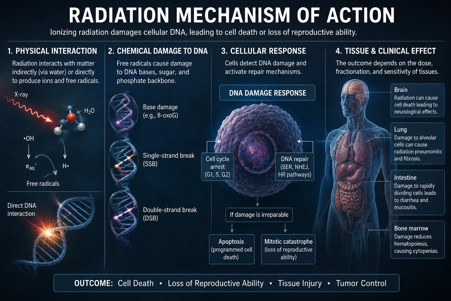
How Radiation Interacts with Cells
Ionizing radiation damages cells through two distinct mechanisms: direct action and indirect action. Understanding the difference is fundamental for the ARRT exam.
Direct Action
In direct action, radiation deposits energy in the DNA molecule itself, causing ionization or excitation and potentially breaking molecular bonds. This is like a bullet hitting its target. For low-LET radiation in water-rich biological material, approximately one-third direct action is a common teaching estimate—not a fixed ratio for every radiation quality or tissue. Direct action becomes relatively more important as LET increases, as with alpha particles.
Indirect Action
In indirect action, radiation ionizes or excites water molecules (H₂O), initiating radiolysis reactions that produce reactive species including the hydrogen radical (H•) and hydroxyl radical (•OH). These are radicals because they contain an unpaired electron; H⁺ and OH⁻ are ions, not the radical notation. The highly reactive •OH can then damage DNA. For low-LET radiation in water-rich tissue, roughly two-thirds indirect action is a useful teaching estimate rather than a universal biological constant.
Cellular Response to Radiation
Not all cells respond to radiation the same way. A cell's radiosensitivity depends on its type, stage in the cell cycle, and metabolic activity.
The Law of Bergonié and Tribondeau (1906)
This foundational law of radiation biology states that cells are most radiosensitive when they have:
- High mitotic activity — they divide rapidly
- Poor differentiation — they are primitive, not yet specialized
- Long mitotic future — they will continue dividing many more times
In other words, the most radiosensitive cells are those that are actively dividing, unspecialized, and have many future divisions ahead. This explains why cancer cells (rapidly dividing) are more radiosensitive than normal tissue — and why radiation therapy works.
Radiosensitivity of Human Tissues
Using the Law of Bergonié and Tribondeau, we can rank human tissues from most to least radiosensitive:
| Radiosensitivity Level | Tissues | Why |
|---|---|---|
| Very High | Hematopoietic stem cells (bone marrow), lymphoid tissue, spermatogonia, intestinal crypt cells | Rapidly dividing, undifferentiated, long mitotic future |
| High | Oocytes, skin stem cells, gastric mucosa, intestinal epithelium | Actively dividing with moderate differentiation |
| Moderate | Lens of eye (epithelium), growing bone and cartilage, vascular endothelium | Some mitotic activity |
| Low | Mature bone and cartilage, salivary glands, liver, kidney, pancreas | Slow or infrequent division, well-differentiated |
| Very Low | Nervous tissue (neurons), muscle, adult brain tissue | Minimal or no mitotic activity, highly differentiated |
Cell Cycle and Radiosensitivity
Cells in different phases of the cell cycle have different radiosensitivity:
- Most radiosensitive: M phase (mitosis) and G₂ phase (pre-mitotic)
- Most resistant: S phase (DNA synthesis) — the cell is already actively repairing DNA during replication
- Intermediate: G₁ phase (pre-synthetic)
M phase is the most radiosensitive because the chromosomes are condensed and visible, making DNA damage more likely to cause cell death during division. This is why rapidly dividing populations like bone marrow and intestinal crypts are hit hardest by radiation.
Cellular Damage: Types of Radiation Injury
Radiation can damage cells in several ways, ranging from repairable to lethal:
| Type of Damage | Description | Outcome |
|---|---|---|
| Sublethal damage | DNA damage that can be repaired if the cell has time between exposures (typically 4–6 hours) | Cell survives; repair is complete |
| Potentially lethal damage | Damage that would kill the cell unless conditions are changed (e.g., less oxygen, delayed division) | Cell may survive under favorable conditions |
| Lethal damage | Irreparable damage — typically double-strand DNA breaks or severe chromosome aberrations | Cell dies (apoptosis or mitotic death) |
Types of Cell Death
Radiation can cause cell death through two main pathways:
- Apoptosis (programmed cell death) — the cell destroys itself in a controlled manner. This can happen within hours of exposure, especially in lymphoid tissues. Apoptosis is an active process — the cell "knows" the damage is too severe and triggers its own shutdown.
- Mitotic death (reproductive death) — the cell attempts to divide but fails because of unrepaired DNA damage. This is the most common form of radiation-induced cell death. The cell may appear functional after exposure but dies when it tries to enter mitosis.
Deterministic vs Stochastic Effects
One of the most important distinctions in radiation biology — and a frequent ARRT exam topic — is the difference between deterministic and stochastic effects.
Deterministic Effects (Tissue Reactions)
Deterministic effects, now commonly called tissue reactions, are characterized by a threshold region: once enough cells are injured, the reaction's incidence and severity increase with dose. Published "thresholds" are practical population estimates—ICRP generally defines them near the dose expected to produce the effect in about 1% of exposed people—not absolute boundaries proving zero effect below one exact number.
- Examples: Skin erythema, epilation (hair loss), cataracts, sterility, desquamation, fibrosis
- Threshold: A practical estimated threshold region is used (often the ~1% incidence dose)
- Severity: Increases with dose — more dose = worse effect
- Mechanism: Cell killing — enough cells are killed that tissue function is impaired
- Latent period: Days to years, depending on dose and tissue
Approximate practical threshold estimates relevant to radiography vary with dose rate, exposed area, follow-up, and individual susceptibility:
- Transient skin erythema: ~2 Gy (2000 mGy) — can appear hours after high-dose fluoroscopy
- Epilation (hair loss): ~3 Gy
- Lens of the eye: ~0.5 Gy for radiation-induced, vision-impairing cataract after acute or protracted exposure (ICRP 118)
- Male sterility (temporary): ~0.15 Gy; permanent at ~3.5–6 Gy
- Female sterility (permanent): ~2.5–6 Gy
Stochastic Effects
For radiation protection, stochastic effects are managed as though they have no threshold dose. Under the LNT model, the probability of an effect increases with dose, but the severity of an effect, if it occurs, is independent of dose. The two categories traditionally taught are:
- Carcinogenesis (radiation-induced cancer)
- Heritable effects (germ-cell changes that could affect descendants; biologically plausible and demonstrated in animals, but not directly established in human populations)
- Threshold: No threshold is assumed for protection; individual risk at very low doses cannot be measured reliably
- Probability: Increases with dose — more dose = higher chance
- Severity: Independent of dose — a radiation-induced cancer is no different from a naturally occurring one
- Mechanism: Mutation of a single cell that survives and reproduces
- Latent period: Years to decades
| Feature | Deterministic | Stochastic |
|---|---|---|
| Threshold? | Practical population threshold estimate, not an absolute zero-effect boundary | No threshold assumed by the LNT protection model |
| Severity vs Dose | Severity increases with dose | Severity is independent of dose |
| Probability vs Dose | Probability increases above threshold | Probability increases with dose |
| Dose-response shape | Sigmoid (S-curve) above threshold | Linear, no threshold (LNT model) |
| Primary mechanism | Cell killing (tissue damage) | DNA mutation in surviving cells |
| Latent period | Hours to years | Years to decades |
| Examples | Skin erythema, cataracts, hair loss | Cancer; potential heritable effects (animal evidence, without direct established human evidence) |
Dose-Response Relationships
Radiation biology uses dose-response models to describe the relationship between radiation dose and biological effect. The two most important models are:
Linear No-Threshold (LNT) Model
The LNT model is the prudent basis for current radiation protection guidelines. It assumes that stochastic cancer risk is proportional to dose without a threshold. At very low doses, epidemiologic data cannot reliably distinguish a small radiation effect from normal cancer variation, so the true shape of the dose-response relationship remains uncertain. LNT is therefore used for planning and optimization—not as proof that a particular tiny dose caused harm or that its numerical risk can be measured precisely.
Threshold Model
Used for tissue reactions. Below the estimated threshold region, the reaction is unlikely to be clinically detectable in a population; above it, incidence and severity rise with dose. The quoted threshold is practical rather than an absolute zero-effect boundary.
You'll also hear about the linear-quadratic model, which is used in radiation therapy to describe cell survival curves at different dose levels. For the ARRT exam, focus on understanding LNT (stochastic) and threshold (deterministic) models.
Factors Affecting Radiosensitivity
Several factors affect how radiosensitive a cell or tissue is:
- Oxygen effect: Well-oxygenated cells are 2–3 times more radiosensitive than hypoxic cells. This is expressed as the oxygen enhancement ratio (OER). It's why radiation therapy tumors with poor blood supply (necrotic centers) are harder to treat.
- LET (Linear Energy Transfer): High-LET radiation (alpha particles, neutrons) causes more damage per unit dose than low-LET radiation (X-rays, gamma rays). Low-LET radiation produces fewer ionizations per unit track length, so the cell has a better chance of repairing the damage.
- RBE (Relative Biological Effectiveness): Compares doses of a reference and test radiation that produce the same specified biological endpoint: RBE = (reference dose) / (test dose). RBE is not one fixed number for a radiation type; it varies with LET, dose, dose rate, tissue, endpoint, and reference beam. Do not equate alpha-particle RBE with 20: wR = 20 is the radiation weighting factor assigned to alpha particles for protection quantities, not a universal measured RBE.
- Dose rate: Lower dose rates allow more time for sublethal damage repair, reducing overall effect. Higher dose rates deliver damage faster than cells can repair it.
- Fractionation: Splitting the total dose into smaller fractions allows normal tissue to repair between exposures while damaging tumor cells more effectively. This is the foundation of radiation therapy.
- Age: Younger organisms are generally more radiosensitive because they have more rapidly dividing cells. Fetuses are the most radiosensitive of all.
Acute Radiation Syndrome (ARS)
Acute Radiation Syndrome (ARS) can follow a high dose of penetrating radiation delivered to all or most of the body in a short time. Routine diagnostic imaging does not meet those exposure conditions. ARS remains important for emergency preparedness and for understanding dose-response relationships.
LD50/60
The LD50/60 is the whole-body dose expected to be lethal to 50% of an exposed population within 60 days. CDC guidance gives an approximate human range of 2.5–5 Gy without medical treatment. It is not one fixed universal dose: outcome varies with dose distribution, radiation quality, age and health, combined injuries, and the availability and timing of supportive care.
Stages of ARS
- Prodromal stage (minutes to hours post-exposure) — nausea, vomiting, diarrhea, fatigue. The speed of onset and severity correlate with dose.
- Latent stage (hours to days) — symptoms temporarily improve. The length of this stage is inversely related to dose.
- Manifest illness stage — symptoms of the specific syndrome appear, depending on which organ system is affected.
- Recovery or death — depending on dose and medical care.
Three Subsyndromes of ARS
| Syndrome | Dose Range | Primary Organ | Outcome |
|---|---|---|---|
| Hematopoietic | Typically ~0.7–10 Gy | Bone marrow | Depression of blood-cell production, with infection and bleeding risk; severity and survival depend strongly on dose and treatment |
| Gastrointestinal | Some GI symptoms from ~6 Gy; full syndrome usually >10 Gy | Intestinal mucosa | Severe diarrhea, fluid loss, infection — often fatal within 1–2 weeks |
| Cardiovascular/CNS | Some symptoms from ~20 Gy; full syndrome usually >50 Gy | Central nervous system | Disorientation, seizures, coma — fatal within hours to days |
Fetal and Embryonic Effects
Radiation effects on the developing embryo and fetus deserve special attention — both because fetal tissue is extremely radiosensitive and because this is a commonly tested ARRT topic.
Effects by Postconception Stage
- Pre-implantation (0–2 weeks): The principal high-dose concern is embryonic death—the classic "all-or-none" teaching pattern—rather than malformation in a surviving embryo.
- Organogenesis (about 2–8 weeks): This is the major period of concern for radiation-induced structural malformations and growth disturbance.
- Early fetal CNS development (8–15 weeks): The brain is most sensitive to radiation-associated severe intellectual disability; sensitivity is lower but remains present at 16–25 weeks.
- Later fetal period: Growth effects and other noncancer tissue reactions become less likely with advancing development, while a potential increase in childhood cancer is considered a stochastic risk throughout pregnancy.
According to CDC guidance, noncancer fetal effects are generally not detectable below about 0.1 Gy fetal dose; severe intellectual disability is of particular concern at substantially higher doses, especially above about 0.5 Gy during the most sensitive CNS period. These fetal doses are far above those from most properly performed diagnostic examinations, but every case still requires justification and optimization.
There is no universal mandatory 10-day rule. Pregnancy screening should follow local law and institutional policy, document menstrual and pregnancy history when the uterus could receive a meaningful dose, and balance fetal protection against the harm of delaying a justified examination. Many settings use a 28-day approach for selected pelvic or lower-abdominal examinations rather than restricting all such imaging to the first 10 days after menstruation. If pregnancy is possible or confirmed, consult the radiologist or designated practitioner and optimize the examination rather than automatically withholding needed care. For more on this, see our complete Radiation Safety Guide.
Practical Applications in Radiography
Understanding radiation biology leads directly to safer practice:
- ALARA makes biological sense — the prudent LNT protection model treats avoidable dose reduction as reducing modeled stochastic risk proportionally, despite uncertainty at very low doses. Our Pediatric Dose Reduction Guide applies these principles specifically to children, who have greater radiosensitivity and longer life expectancy to manifest stochastic effects.
- Collimation protects more radiosensitive organs outside the field of interest — think ovaries, testes, and thyroid during a chest X-ray.
- Gonadal shielding protects reproductive cells, which are among the most radiosensitive in the body. (See our Patient Shielding Guide for current recommendations.)
- Proper positioning to avoid repeats is the single most effective dose reduction strategy — every repeat doubles the dose for that examination.
- Fluoroscopy time and dose awareness — around 2 Gy is a practical threshold estimate for transient erythema after localized skin exposure, so substantial cumulative skin dose requires monitoring and follow-up. Our Fluoroscopy Procedures and Safety Guide covers dose monitoring in detail.
- Pediatric patients require lower technique factors and special attention — children have more rapidly dividing cells and a longer lifetime for stochastic effects to develop.
ARRT Exam Review: Key Points to Remember
Here's a condensed review of the highest-yield radiation biology concepts for the ARRT registry:
Direct vs Indirect
For low-LET radiation in water-rich tissue, ~⅔ indirect and ~⅓ direct action is a teaching estimate. Water radiolysis produces H• and •OH radicals; oxygen can fix radical-mediated damage.
Bergonié & Tribondeau
Cells are most radiosensitive when they are rapidly dividing, undifferentiated, and have a long mitotic future.
Deterministic vs Stochastic
Tissue reactions use practical population threshold estimates; severity ↑ with dose. For stochastic risk, LNT prudently assumes no threshold and probability ↑ with dose.
Most Radiosensitive
Bone marrow, lymphoid tissue, spermatogonia, intestinal crypts. Least: nerve cells, muscle, adult brain.
Cell Cycle
M phase = most radiosensitive. S phase = most resistant. Rapidly dividing populations get hit hardest.
Fetal Effects
Organogenesis (2–8 weeks postconception) is the structural-malformation period. Severe intellectual-disability sensitivity is greatest at 8–15 weeks and lower at 16–25 weeks.
For a broader review of physics fundamentals, revisit our X-Ray Physics Made Simple guide — radiation biology is the bridge between physics and clinical practice.
Test Your Radiation Biology Knowledge
Try these ARRT-style multiple choice questions based on this article. Click an option to check your answer — correct answers turn green, wrong ones turn red.
Skin erythema is a tissue reaction. About 2 Gy is a practical population threshold estimate for transient erythema—not an absolute boundary—and severity rises as dose increases above the threshold region. A localized skin reaction is distinct from ARS, which requires a large acute exposure of most or all of the body. Cancer risk is handled with the LNT model and would not present as erythema two days later; heritable effects concern germ cells, not a local skin response.
Hematopoietic stem cells are rapidly dividing, poorly differentiated, and have a long mitotic future — all three criteria from Bergonié and Tribondeau that make cells highly radiosensitive. Mature neurons and cardiac muscle cells rarely divide (highly differentiated), and liver cells divide only when needed. This is why bone marrow suppression is the earliest sign of significant radiation exposure.
For low-LET radiation in water-rich tissue, indirect action accounts for approximately two-thirds (67%) in a commonly used teaching estimate. Water radiolysis generates reactive species, especially the hydroxyl radical •OH, which can attack DNA; H• and •OH are radicals, whereas H⁺ and OH⁻ are ions. The remaining one-third is attributed to direct energy deposition in DNA. The ratio is not universal: it changes with radiation quality, LET, and biological conditions.
Two key factors make children more radiosensitive for stochastic effects: (1) they have more rapidly dividing cells (Law of Bergonié and Tribondeau), making DNA damage more likely to become permanent, and (2) they have a longer remaining lifetime for radiation-induced cancers to develop (latency period for solid tumors is 10–20+ years). This is why pediatric protocols emphasize lower kVp, reduced mAs, and rigorous adherence to ALARA — covered in our Pediatric Dose Reduction Strategies guide.
M phase (mitosis) is the most radiosensitive phase of the cell cycle because the chromosomes are condensed into visible structures and the cell is actively dividing. Any DNA damage is more likely to result in mitotic death (reproductive death) because the cell cannot properly segregate damaged chromosomes. The S phase is the most radioresistant because the cell's DNA repair mechanisms are already active during replication. This is why rapidly dividing tissues — bone marrow, intestinal crypts, and tumors — are most affected by radiation.