X-Ray Production: How X-Rays Are Made
Every X-ray image starts in one place: the X-ray tube. Understanding exactly how X-rays are produced — from the moment the exposure button is pressed to the instant photons exit the tube port — is the foundation of radiographic physics. This article breaks down the two mechanisms of X-ray production, the components involved, and the spectrum of energies that result.
Key Concept
X-rays are produced by rapid deceleration of electrons. When high-speed electrons strike the anode target, they lose kinetic energy — that lost energy is emitted as X-ray photons. Only about 1% of the energy becomes X-rays; the other 99% becomes heat.
The X-Ray Tube: A Quick Overview
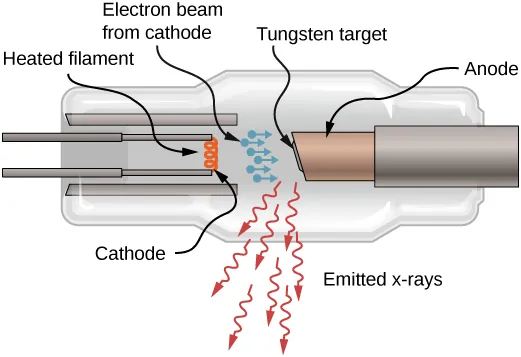
Before we dive into how X-rays are produced, let's review the hardware that makes it happen. The X-ray tube is a vacuum-sealed glass or metal envelope containing two main electrodes:
Cathode (−)
Contains the filament (usually tungsten wire) that produces electrons via thermionic emission when heated. Also has a focusing cup to direct the electron stream toward the anode.
Anode (+)
Target surface (tungsten or tungsten-rhenium alloy) where electrons impact. Rotating anodes dissipate heat across a larger area. The angle of the target face affects focal spot size.
Envelope & Housing
The vacuum glass envelope allows electrons to travel unimpeded. The lead-lined housing contains radiation and the oil bath provides electrical insulation and heat dissipation.
The Two Mechanisms of X-Ray Production
When electrons strike the anode, X-rays are produced through two distinct physical processes. Both happen simultaneously inside the tube. Understanding the difference is essential for mastering X-ray physics and image optimization.
1. Bremsstrahlung Radiation ("Braking Radiation")
Bremsstrahlung (pronounced brehm-strah-lung) is German for "braking radiation." It accounts for approximately 80-90% of X-ray photons produced in a diagnostic X-ray tube operating above 70 kVp.
How It Works
When a high-speed electron passes near the nucleus of a tungsten atom:
- The electron is attracted by the strong positive charge of the nucleus
- Its path is deflected (bent), causing it to slow down
- The lost kinetic energy is emitted as an X-ray photon
The closer the electron passes to the nucleus, the greater the deflection and the more energy is released as an X-ray photon. This creates a continuous spectrum of X-ray energies — from very low energy (distant pass) up to the maximum kVp setting (direct hit).
ARRT Tip
The maximum energy of a Bremsstrahlung photon equals the tube voltage in kVp. At 80 kVp, the maximum photon energy is 80 keV — no X-rays exist above this energy. The average energy of the beam is roughly 30-40% of kVp.
2. Characteristic Radiation
Characteristic radiation produces X-ray photons at specific, discrete energies determined by the target material. For tungsten, these are approximately 59-69 keV.
How It Works
- An incoming electron has enough energy to eject an inner-shell electron (K-shell) from a tungsten atom
- This leaves a "hole" or vacancy in the K-shell
- An electron from an outer shell (L-shell, M-shell) drops down to fill the vacancy
- The energy difference between shells is released as an X-ray photon with a specific energy
Characteristic Radiation Energies for Tungsten
| Transition | Energy (keV) | Common Name |
|---|---|---|
| L-shell → K-shell | 59.3 | Kα (K-alpha) |
| M-shell → K-shell | 67.2 | Kβ (K-beta) |
When Does Characteristic Radiation Occur?
Characteristic radiation only occurs when the kVp exceeds the K-shell binding energy of the target material. For tungsten, this threshold is 69.5 keV. Below this kVp, all X-rays are Bremsstrahlung. Above it, characteristic K-shell X-rays appear as sharp peaks in the emission spectrum.
The X-Ray Emission Spectrum
The X-ray emission spectrum is a graph plotting the number of photons (y-axis) against their energy in keV (x-axis). It tells you everything about the quality of the X-ray beam.
Reading the Spectrum
▸ Smooth curve = Bremsstrahlung continuum. ▸ Orange spikes = Characteristic radiation peaks (Kα and Kβ).
The spectrum reveals three important facts:
- No X-rays at zero energy — low-energy photons are filtered out by the tube housing and added filtration
- The curve rises gradually — peaks at about 30-40% of kVp, then drops to zero at the maximum kVp setting
- Sharp spikes — characteristic radiation peaks superimposed on the continuous Bremsstrahlung curve (only present above 69.5 kVp for tungsten)
What Changes the Spectrum?
| Adjustment | Effect on Spectrum |
|---|---|
| Increase kVp | Shifts the entire curve to the right (higher energies). Maximum energy = new kVp. Higher average energy = more penetrating beam. |
| Increase mAs | Amplifies the curve upward (more photons at every energy). Does not change beam quality. |
| Add filtration | Removes low-energy photons from the left side of the curve. "Hardens" the beam — higher average energy, reduced patient dose. |
| Change target angle | Steeper angle = more heel effect. Softer beam on the anode side of the field. |
Why 99% Heat?
Only about 1% of electron kinetic energy converts to X-rays. The remaining 99% becomes heat in the anode. This is why:
- Tungsten is used for the target — it has the highest melting point of any metal (3,422°C)
- Rotating anodes spin at 3,000-10,000 RPM to spread heat across a larger track
- Modern tubes use a tungsten-rhenium alloy target bonded to a molybdenum or graphite backing for better heat capacity
- The tube housing is surrounded by oil that absorbs and dissipates heat
Heat vs. X-Rays — A Useful Analogy
Think of the X-ray tube like an incandescent light bulb: the filament gets hot and produces light (X-rays) as a byproduct. But instead of visible light, the "glow" from the anode is in the X-ray part of the electromagnetic spectrum. You can explore the X-Ray modality page for more on clinical applications.
Putting It All Together: The Full Production Chain
- Filament current heats the cathode filament to ~2,200°C (thermionic emission releases electrons)
- Tube voltage (kVp) accelerates electrons from cathode to anode at nearly 50% the speed of light
- Electron impact — electrons strike the rotating tungsten anode target
- Bremsstrahlung — electrons deflected by nuclei produce ~80-90% of X-rays (continuous spectrum)
- Characteristic — if kVp > 69.5, K-shell interactions produce discrete energy peaks (Kα, Kβ)
- Filtration — inherent + added filtration removes low-energy photons (patient dose reduction)
- Collimation — lead shutters shape the beam to the anatomy of interest
- Exit port — the useful X-ray beam emerges through the tube housing window
Key Takeaways for the Registry
- Bremsstrahlung = continuous spectrum, accounts for most photons, occurs at all kVp levels
- Characteristic radiation = discrete energies, only above 69.5 kVp for tungsten
- Maximum photon energy = kVp setting (at 90 kVp, max photon is 90 keV)
- Average photon energy ≈ 30-40% of kVp (varies with filtration)
- 99% heat, 1% X-rays — always remember the efficiency ratio
- Filtration hardens the beam by removing low-energy photons
For more on how these physics principles translate to image quality, read our guide to X-Ray Physics Made Simple: kVp, mAs, Density, and Contrast.