CT Scan — Computed Tomography
CT combines a rotating X-ray tube with a bank of detectors to acquire cross-sectional (axial) images of the body. Computer reconstruction creates a full 3D volumetric dataset from hundreds of projections.
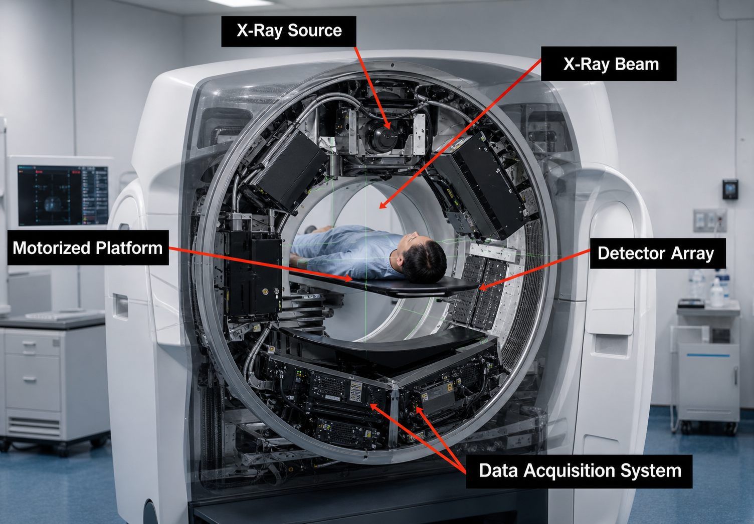
This CT scanner is shown with the outer housing removed to reveal the rotating internal assembly.
CT Gantry Components
The gantry houses the rotating X-ray tube and detector assembly. Below are the key internal components shown in the cutaway image.
Rotating anode (up to 200mm), 80–140 kVp. Engineered for continuous high-output rotation. Dual-source CT uses two tubes 90° apart for cardiac imaging. Tube heat capacity (MHU) is a critical spec.
Two sets: pre-patient (near tube) define slice thickness and irradiated volume; post-patient (near detectors) reduce scatter reaching the detector rows, improving image contrast.
A 30–60° fan beam spans the patient from tube to curved detector array. Each rotation angle records a projection of tissue attenuation — hundreds to thousands per rotation are reconstructed into axial slices.
Circular electrical contacts enabling continuous gantry rotation — no cables to rewind. Enables helical CT (patient moves while gantry rotates). Modern systems use optical slip rings for high-speed data transmission.
Cylindrical opening (70 cm standard, 80–90 cm wide-bore). Contains rotating tube/detector assembly, lasers, cooling, and slip-ring connections. The isocenter (beam intersection point) defines optimal resolution and dose.
Curved solid-state scintillator + photodiode array. 64-slice = 64 active rows; 320-slice covers 16cm in a single rotation (whole-heart cardiac CT). Photon-counting detectors measure individual photon energies for spectral imaging at lower dose.
Carbon-fiber couch advances smoothly during helical scanning. Speed linked to pitch and rotation time. Accuracy ±0.25 mm for reproducible positioning. Weight limit 200–250 kg.
Key Concepts
- Reconstruction: Filtered back projection (FBP) or iterative reconstruction algorithms calculate tissue attenuation at each voxel from hundreds of angular projections
- Isotropic voxels: 64+ slice MDCT produces equal x/y/z resolution — reconstruct in any plane (axial, coronal, sagittal) without quality loss
- Pitch: Table advance per rotation ÷ total beam width. Pitch >1 = faster, less dose; Pitch <1 = overlap, more dose
- Helical / Spiral CT: Continuous rotation + continuous table movement creates a helical data acquisition through the body volume
- Kernels: Soft-tissue kernel (smooth) for abdomen/brain; sharp/bone kernel for high-contrast structures like cortical bone
Hounsfield Units (HU)
CT quantifies tissue density using the Hounsfield scale. Each voxel is assigned an HU value based on its X-ray attenuation relative to water. This standardized scale allows tissue characterization across all CT scanners worldwide.
Click a tissue below — the orange marker will jump to its position on the scale.
Click a Tissue to See Its HU Range
Window & Level (W/L)
CT displays a narrow range of HU values at a time. Window Width (WW) sets the range; Window Level (WL/C) sets the center. Only this is displayed in grayscale — outside the window appears black or white.
| Window Preset | Width (HU) | Level (HU) |
|---|---|---|
| Brain (soft) | 80 | 40 |
| Subdural | 200 | 60 |
| Stroke/Bone | 1500–2000 | 300–400 |
| Lung | 1500 | −600 |
| Abdomen | 350 | 40 |
| Liver | 150 | 70 |
| Bone | 2000 | 300 |
CT Generations
CT technology has evolved through five generations, each improving speed, resolution, and dose efficiency.
Pencil Beam · Translate-Rotate
Single pencil beam and 1–2 detectors. Tube and detector translate across the patient, then rotate 1°. Slow, precise, and mainly used for early head imaging.
Select a generation card to animate how the source, beam, and detector geometry acquire projection data.

🔴 Pencil Beam · Translate-Rotate
Single pencil beam and 1–2 detectors. Tube and detector translate across the patient, then rotate 1°. Scan time: ~4.5 min/slice.

🔶 Narrow Fan Beam · Translate-Rotate
Narrow fan beam with multiple detectors (3–30). Still translate-rotate but larger rotation increments. Scan time: ~20 s/slice.

🟠 Wide Fan Beam · Rotate-Rotate
Wide fan beam (30–60°) with hundreds of detectors in an arc, both rotating together. No translation. Scan time: <1 s/rotation.

🟡 Fixed Detector Ring
Stationary ring of 600–4800 detectors. Only the X-ray tube rotates. Eliminates ring artifacts but expensive — each detector needs its own electronics.
🟣 Electron Beam CT (EBCT)
No rotating tube. Electron beam is steered electromagnetically around a tungsten target ring. Ultra-fast: 50–100ms per slice.
CT Scan Parameters
Understanding these parameters helps optimize image quality while minimizing radiation dose — a key skill for every radiographer.
| Parameter | Definition | Effect on Image | Effect on Dose |
|---|---|---|---|
| kVp | Peak tube voltage (80–140 kVp) | ↑kVp = less subject contrast, better penetration | ↑kVp = ↑dose if mAs is unchanged |
| mAs | Tube current × time | ↑mAs = less noise (↑SNR) | ↑mAs = ↑dose (linear) |
| Pitch | Table advance ÷ beam width | ↑pitch = more noise | ↑pitch = ↓dose |
| Slice Thickness | Reconstructed slice width (mm) | Thinner = better resolution, more noise | No direct change |
| FOV | Display reconstruction circle | Smaller = better pixel size | No direct change |
| Kernel | Reconstruction filter | Soft: smooth; Sharp: high res, noisy | No change |
| CTDIvol | CT Dose Index — dose per rotation | — | Primary dose metric |
| DLP | CTDIvol × scan length | — | Total dose metric |
Dose Optimization (ALARA): CT delivers significantly more dose than plain radiography. Use automatic exposure control (AEC), iterative reconstruction, and lowest acceptable kVp/mAs for each indication. CT of the abdomen/pelvis ≈ 300–700 chest X-rays in effective dose.
Common CT Artifacts
Recognising artifacts is essential — they can mimic pathology or hide real findings. Each one has a specific cause and a specific fix.
Radiographer tip: Most artifacts are recognised by their pattern, not by looking at the patient — check for symmetry around the isocentre, alignment with dense structures, or correlation with respiratory phase before blaming pathology.
Clinical Applications
CT is the workhorse of emergency and complex diagnostic imaging, offering rapid whole-body assessment.
🚑 Trauma
Whole-body trauma CT (pan-scan) in seconds. Detects intracranial hemorrhage, pneumothorax, solid organ injuries, and vascular disruption. Standard of care in major trauma.
🫀 CT Angiography
CTA of brain, carotids, coronary, aorta, pulmonary, and peripheral arteries. Replaced conventional angiography for most diagnostic purposes. PE protocol for pulmonary embolism.
🎗️ Oncology
Tumor staging, treatment planning, response assessment. CT-guided biopsy and ablation. PET/CT combines metabolic and anatomical information for oncology staging.
🧠 Neuroimaging
Acute stroke triage (NCCT + CTA + perfusion), intracranial hemorrhage, skull fractures, and hydrocephalus. First-line in acute neurological presentation.
🫁 Chest & Abdomen
HRCT for interstitial lung disease, nodule characterization (LDCT lung cancer screening), appendicitis, diverticulitis, bowel obstruction, and renal calculi.
🦷 Dental / CBCT
Cone-beam CT (CBCT) provides low-dose 3D dental and maxillofacial imaging for implant planning, orthodontics, and TMJ assessment.
Inside the Nucleus
Nuclear Medicine — imaging the body's metabolic activity with radioactive tracers.
Explore Nuclear Medicine →