History of Radiology
From a glowing fluorescent tube in 1895 to AI-assisted diagnostics today — explore the remarkable century-long journey that built modern medical imaging.
Milestones in Medical Imaging
Click any milestone to read more about that discovery or innovation.
Röntgen Discovers X-Rays
Wilhelm Conrad Röntgen accidentally discovers a new type of ray while experimenting with cathode ray tubes in Würzburg, Germany.
Röntgen noticed that a fluorescent screen nearby began to glow even though it was not in the direct path of the cathode ray tube. He realized a new, unknown type of ray was passing through the cardboard shield. He named them "X-rays" — X for unknown. Within weeks he produced the first medical X-ray image, of his wife Anna Bertha's hand, revealing the bones and her ring. The discovery earned him the first Nobel Prize in Physics in 1901.
First Clinical X-Ray Images
Within months of Röntgen's announcement, physicians worldwide began using X-rays for diagnosis — locating bullets, fractures, and foreign bodies.
In January 1896, just 6 weeks after Röntgen's paper, Dr. John Hall-Edwards in Birmingham used X-rays to guide surgery on a patient's hand. By mid-1896, X-ray equipment was being manufactured commercially. Early machines had no radiation protection, leading to serious radiation injuries among early users.
Marie Curie & Radioactivity
Marie and Pierre Curie discover polonium and radium, laying the groundwork for nuclear medicine and radiotherapy.
Marie Curie coined the term "radioactivity" and discovered that uranium emitted rays that could ionize air. In 1898 she discovered polonium and radium. During World War I, she developed mobile X-ray units (called "petites Curies") that brought X-ray diagnostics to battlefield surgery. Curie won two Nobel Prizes — in Physics (1903) and Chemistry (1911).
Coolidge Tube Invented
William Coolidge develops the hot-cathode X-ray tube, dramatically improving X-ray output, reliability, and control.
The Coolidge tube replaced the unreliable gas tube with a tungsten filament heated to release electrons. This allowed for controlled kVp and mA settings — the same basic principles used in modern X-ray tubes today. Coolidge's design enabled consistent, high-quality radiographic images and is considered one of the most important engineering advances in radiology.
Fluoroscopy & Contrast Media
Fluoroscopic screening allows real-time X-ray viewing. Barium sulfate and iodinated contrast agents expand diagnostic capability to soft-tissue structures.
Fluoroscopy was initially used without any radiation protection, causing many injuries to practitioners. The development of the image intensifier in the 1950s made fluoroscopy safer and more practical. Contrast media like barium sulfate (for GI tract) and iodinated agents (for blood vessels and kidneys) allowed structures invisible on plain films to be visualized.
Birth of Nuclear Medicine
The use of radioactive isotopes for diagnosis expands after World War II. Iodine-131 is first used to treat thyroid disease in 1946.
The Manhattan Project's nuclear research led to the production of artificial radioisotopes in nuclear reactors. Physicians quickly found medical uses — I-131 for thyroid imaging and therapy, Tc-99m for bone and organ scanning. Hal Anger developed the scintillation (gamma) camera in 1958, enabling the imaging of radioisotope distribution throughout the body.
Diagnostic Ultrasound Emerges
Using sonar principles adapted from naval technology, physicians develop diagnostic ultrasound for real-time soft-tissue imaging.
Dr. Ian Donald in Glasgow pioneered the use of ultrasound in obstetrics in the late 1950s, producing the first obstetric ultrasound images. Early scanners used A-mode (amplitude) and later B-mode (brightness) imaging. The development of real-time B-mode scanners in the 1970s and Doppler techniques transformed prenatal care and abdominal diagnostics.
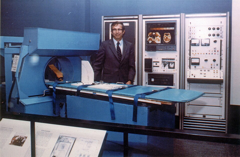
First CT Scanner (EMI Mark 1)
Godfrey Hounsfield and Allan Cormack develop the first practical computed tomography scanner. The first clinical scan is performed in 1971.
Hounsfield, an engineer at EMI Ltd., conceived of reconstructing cross-sectional images from multiple X-ray projections. The first clinical brain CT scan was performed at Atkinson Morley's Hospital, London, on October 1, 1971. It revealed a frontal lobe tumor. The scanner took several minutes to acquire data and hours to reconstruct an image. Hounsfield and Cormack shared the 1979 Nobel Prize in Physiology or Medicine.
MRI: From NMR to Imaging
Paul Lauterbur and Peter Mansfield develop the mathematical framework for magnetic resonance imaging, producing the first MR images of living tissue.
Nuclear magnetic resonance (NMR) had been used in chemistry labs for decades. Paul Lauterbur's key insight (1973) was using magnetic field gradients to encode spatial information, enabling image reconstruction. Peter Mansfield developed fast imaging techniques including echo-planar imaging (EPI). The first human MRI was performed in 1977 by Raymond Damadian. Lauterbur and Mansfield shared the 2003 Nobel Prize in Physiology or Medicine.
PET Scanning & Digital Radiography
Positron emission tomography (PET) enables metabolic imaging; digital radiography (CR then DR) replaces film-screen systems.
PET scanning uses positron-emitting isotopes (like F-18 FDG) to image metabolic activity — invaluable in oncology and neurology. Computed radiography (CR), using photostimulable phosphor plates, was introduced commercially in 1983. Direct digital radiography (DR) flat-panel detectors arrived in the 1990s, dramatically reducing radiation dose while improving image quality.
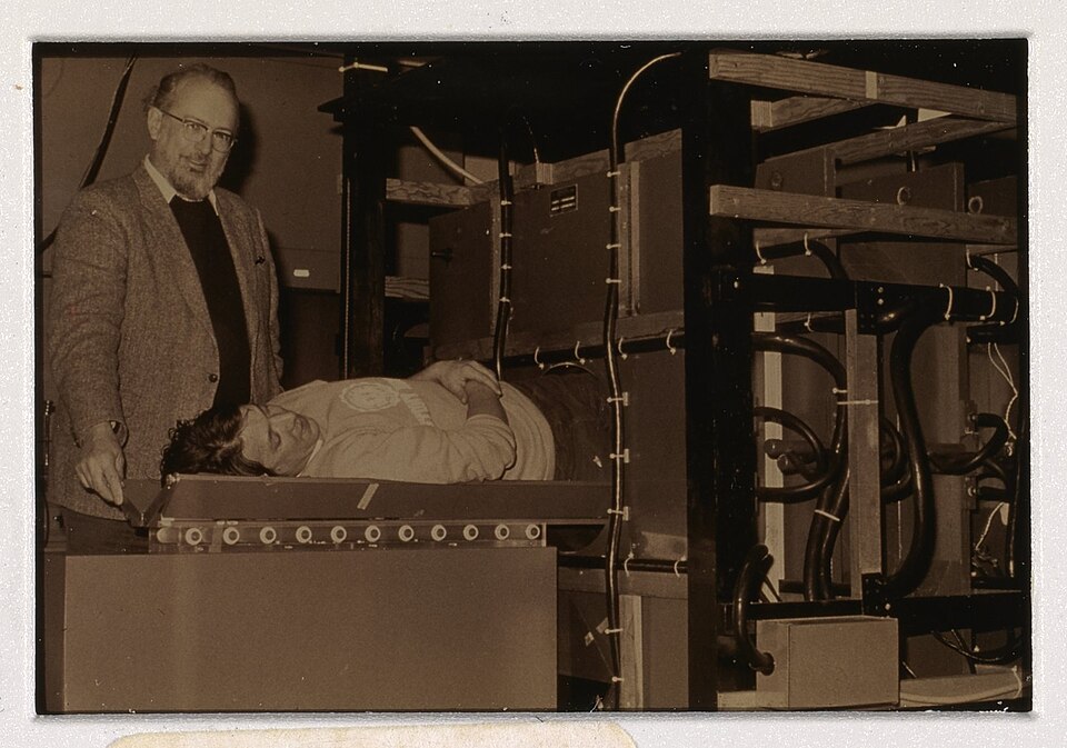
Multidetector CT & 3D Imaging
64-slice and beyond MDCT scanners provide isotropic voxels, enabling 3D reconstruction, CT angiography, and cardiac imaging in seconds.
The introduction of 4-slice (1998), 16-slice (2002), and 64-slice (2004) MDCT revolutionized radiology. Sub-millimeter isotropic voxels allowed 3D multiplanar reconstructions in any plane. CT coronary angiography became possible, and CT pulmonary angiography replaced conventional pulmonary angiography. Dual-energy CT allows material decomposition and virtual non-contrast imaging.
PET/MRI, Spectral CT & PACS
Hybrid scanners combine functional and anatomical imaging; PACS and teleradiology transform workflow and access to images worldwide.
PET/CT (2001) and later PET/MRI (2010s) provided simultaneous anatomical and metabolic imaging. Spectral/photon-counting CT can characterize tissue composition. Picture Archiving and Communication Systems (PACS) digitized the entire radiology workflow, enabling instant access, remote reading, and AI integration. By 2020, radiology was nearly entirely digital.
AI & Deep Learning in Radiology
Artificial intelligence assists with image interpretation, anomaly detection, dose optimization, and workflow automation in radiology departments worldwide.
Deep learning convolutional neural networks (CNNs) now match radiologist performance in specific tasks like chest X-ray triage, mammography screening, and intracranial hemorrhage detection. AI tools integrate into PACS workflows to flag critical findings. Photon-counting detector CT (FDA approved 2021) provides unprecedented low-dose, high-resolution imaging with spectral capabilities.
History image credits
- Anna Bertha Röntgen hand X-ray
- Early X-ray procedure
- Pitchblende uranium ore
- Coolidge X-ray tube
- Fluoroscopy procedure 1909
- Meath gamma camera
- 1985 obstetric ultrasound photo
- EMI 1010 CT scanner
- MRI Scanner Mark One
- ECAT Exact HR PET scanner
- Siemens Somatom Sensation 64 CT
- Radiologist working with modern IT system
- IBM Medical Sieve
People Who Changed Medicine
The scientists and physicians whose discoveries built the field of diagnostic imaging.
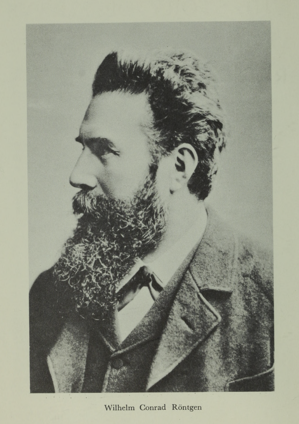
German physicist who accidentally discovered X-rays on November 8, 1895. Won the first Nobel Prize in Physics in 1901.
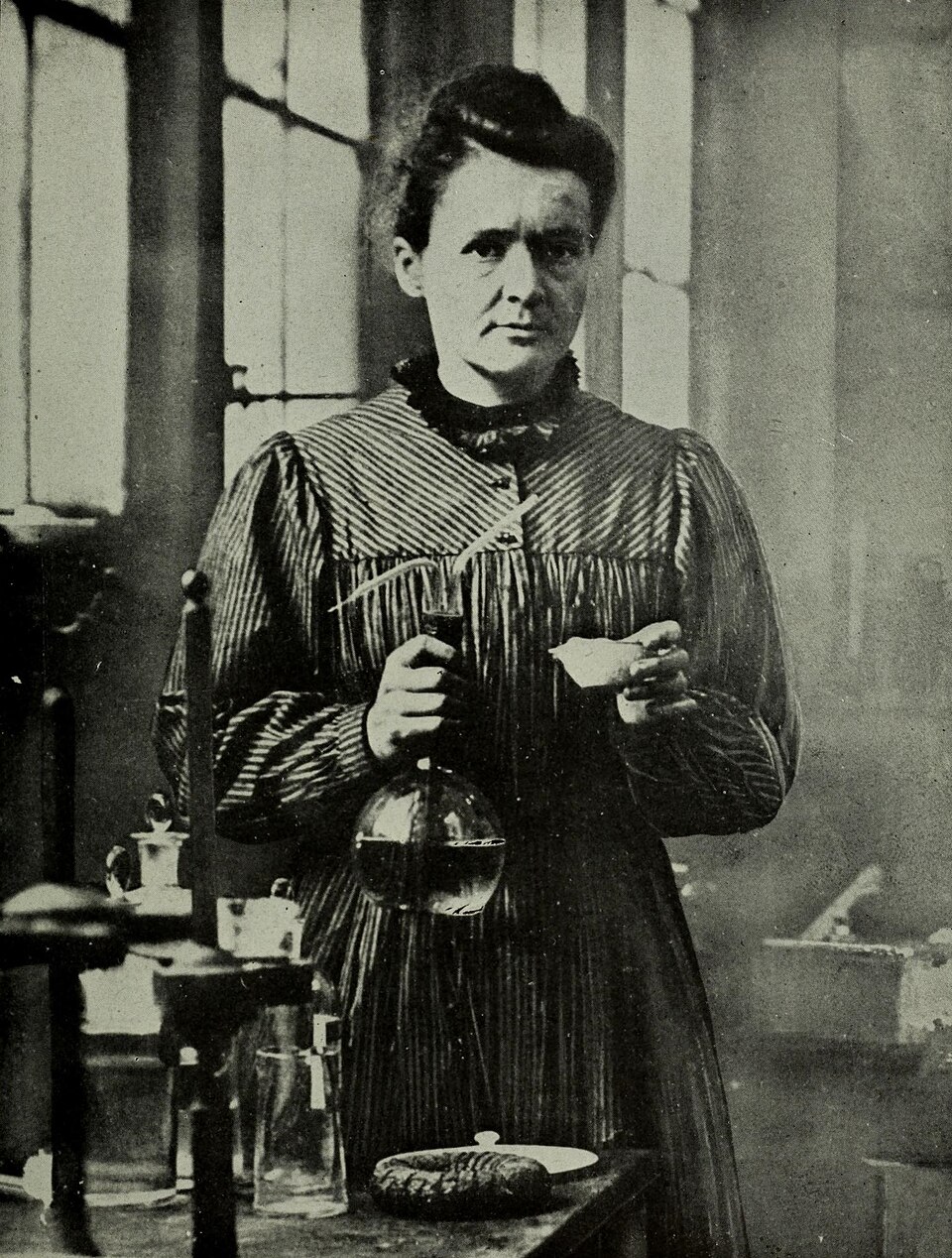
Pioneered research on radioactivity; discovered polonium and radium. Developed mobile X-ray units used in WWI. Two-time Nobel laureate.
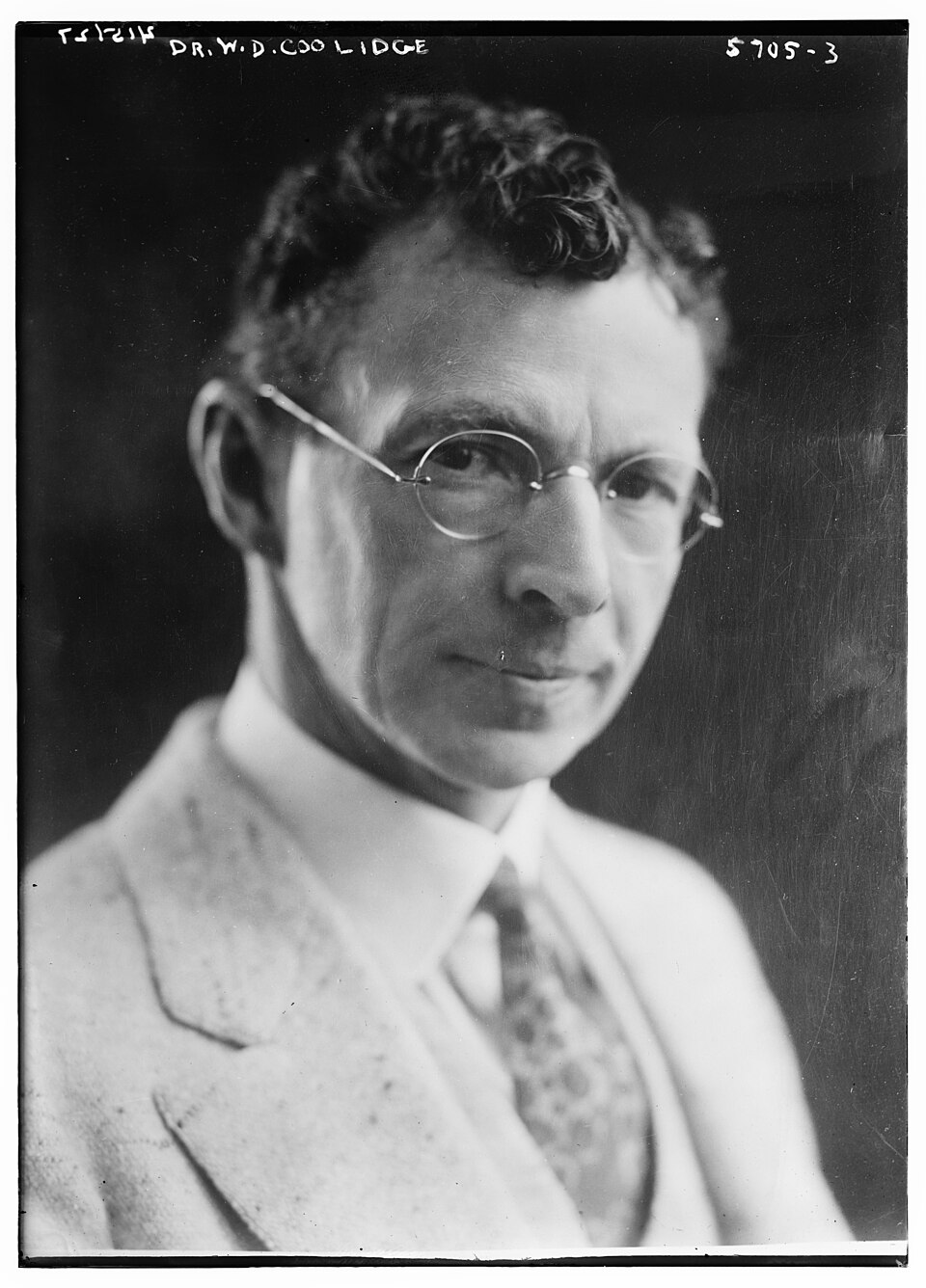
GE physicist who invented the hot-cathode (Coolidge) X-ray tube, the design foundation of all modern X-ray tubes.
EMI engineer who invented computed tomography. The Hounsfield unit (HU) for tissue density is named in his honor. Nobel Prize 1979.
Physicist who developed the mathematical methods that made computer-assisted tomography possible. Shared the 1979 Nobel Prize with Hounsfield.
Chemist who developed the concept of using magnetic gradients to create spatial MRI images. Shared the 2003 Nobel Prize in Medicine.
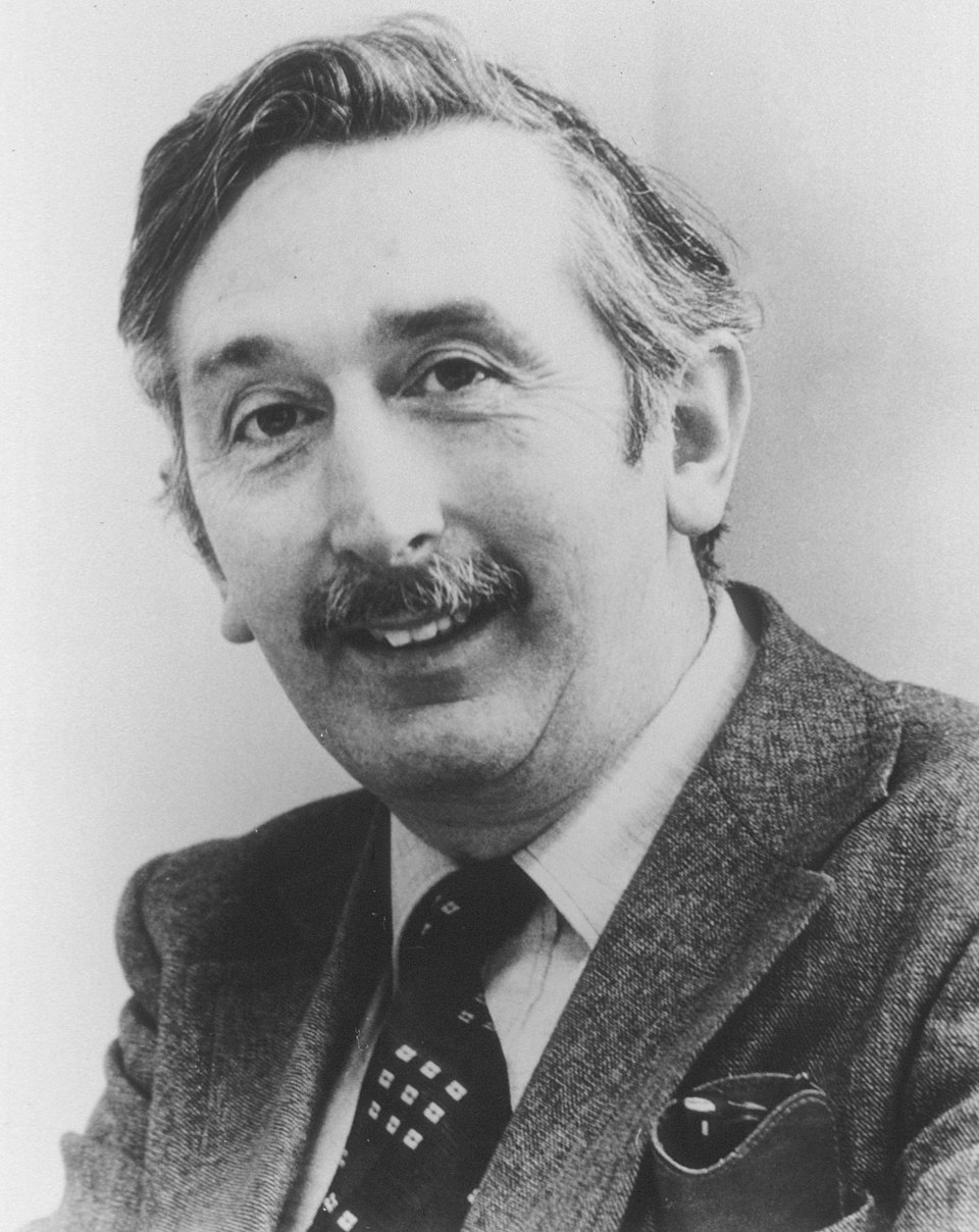
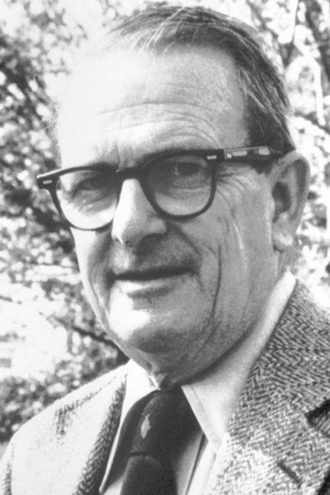
Developed the ACTA scanner, the first whole-body CT system, expanding CT beyond head imaging and pushing medical image reconstruction forward.
Pioneer portrait credits
_machine_-_DPLA_-_4114afc3fdfcbb6da5706c8fe95c480d_(page_2).jpg)
Key takeaway for students: Every modality you'll learn in your program was once a brand-new idea that had to overcome skepticism. The history of radiology is also the history of scientific courage and curiosity — qualities every radiographer carries forward.
Ready to Learn the Physics?
Start with X-Ray — the modality that started it all, and the foundation for CT and fluoroscopy.
Explore X-Ray →