⚡ Modality 1

Diagnostic Radiography

The foundation of medical imaging, utilizing high-energy electromagnetic radiation to visualize the internal structures of the body. X-rays remain the most frequent radiographic procedure, essential for skeletal, thoracic, and emergency imaging.

1895
Discovered
Ionizing
Radiation
2D
Projection
W
Tungsten Target

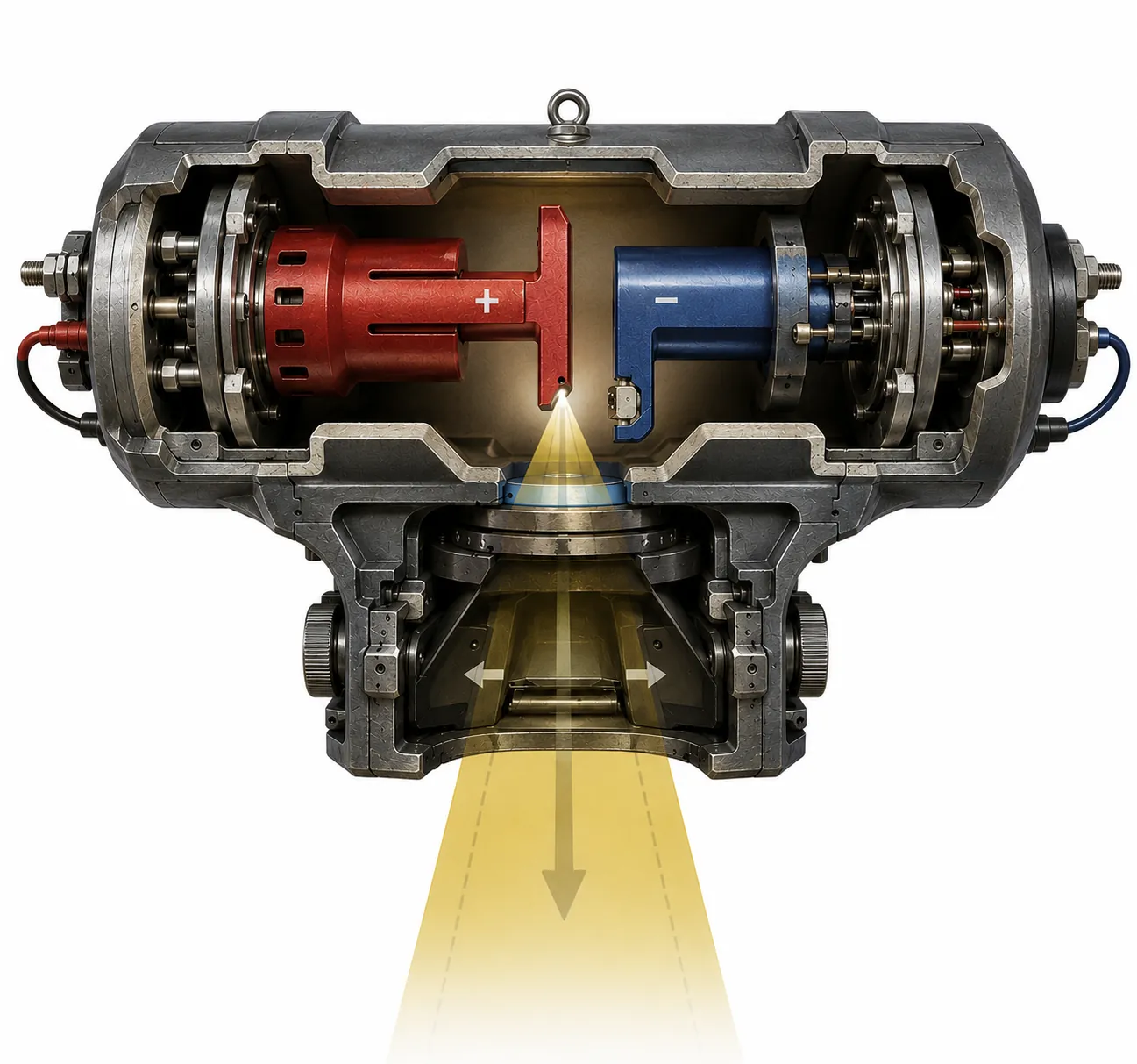
The Physics of X-Rays

X-rays are electromagnetic radiation with wavelengths of 0.01–10 nm, occupying the spectrum between UV and gamma rays. In diagnostic radiology, they are produced artificially using an X-ray tube.

X-rays are generated by two mechanisms inside the tube: Bremsstrahlung radiation (braking radiation — ~80–90% of output) and characteristic radiation (~10–20%).

  • Bremsstrahlung: electrons decelerate near the tungsten nucleus, releasing energy as X-ray photons across a spectrum of energies
  • Characteristic: incident electrons knock inner-shell electrons out of tungsten atoms; outer-shell electrons fill the gap, emitting photons of specific (characteristic) energy
  • X-rays travel at the speed of light (3×10⁸ m/s) and carry no mass or charge
  • Higher kVp → higher energy photons → more penetrating beam → lower contrast
  • Higher mAs → more photons → more exposure → less noise (quantum mottle)
Electromagnetic Spectrum
Radio Microwave Infrared Visible UV X-RAYS 0.01–10 nm Gamma Long λ / Low E Short λ / High E

Tap the tube components, then switch the atom lens between X-ray production in tungsten and photon interactions in tissue.

Photorealistic X-Ray Tube Cutaway
Exposure running

Bremsstrahlung inside tungsten

incident e-
x-ray photon
mostly heat
incident photon
photoelectron
characteristic xray
incident photon
scattered photon
compton electron

An incident tube electron passes close to the positive tungsten nucleus, is deflected and slowed, and the lost kinetic energy leaves as an X-ray photon. Most electron energy still becomes heat in the target.

X-Ray Tube Components

Click on a component in the diagram to learn about it.

  • Typical tube voltage: 40–150 kVp diagnostic; up to 25 kV mammography
  • Tungsten (W, Z=74) is used as the anode target due to its high atomic number and high melting point (3,422°C)
  • Only ~1% of electron energy becomes X-rays; 99% becomes heat
  • Rotating anodes spread heat over a larger focal track area
1

Filament heating releases electrons by thermionic emission.

2

kVp accelerates those electrons across the evacuated glass tube.

3

Electrons strike the rotating tungsten disc, spreading heat around the focal track while producing X-ray photons.

4

Useful photons exit through the window, pass through anatomy, and form contrast at the detector.

How X-Rays Interact with Matter

When an X-ray beam enters the body, photons interact with tissue in five main ways. The dominant interactions in diagnostic radiology are photoelectric absorption and Compton scattering.

🎯

Photoelectric Absorption

Low-energy photon absorption by an inner-shell electron, causing ionization and contrast.

Dominant < 80 kVp
View Interaction

A low-energy X-ray photon transfers all its energy to an inner-shell electron (usually K-shell). The photon disappears, and the electron is ejected as a "photoelectron".

Low Energy Photon Photoelectron

Clinical: Essential for producing image contrast (bone appearing white).

↗️

Compton Scattering

Photon interaction with an outer-shell electron, creating scatter and reduced contrast.

Dominant 80-150 kVp
View Interaction

An intermediate-energy X-ray photon strikes a loosely bound outer-shell electron. The photon is "scattered" with reduced energy, creating occupational dose.

Clinical: The primary source of scatter radiation and film fog.

➡️

Coherent Scatter

Very low-energy interaction causing atom oscillation with no ionization.

Below 30 kVp
View Interaction

The photon causes atom vibration, and a new photon of same energy is emitted in a slightly different direction. Minimal impact on imaging.

Clinical: Minor contributor, usually ignored in diagnostic kVp ranges.

⚛️

Pair Production

High-energy photon conversion into an electron-positron pair near the nucleus.

Above 1.02 MeV
View Interaction

High-energy photon interacts with the nuclear force field and is converted into a negatively charged electron and a positively charged positron.

Electron (-) Positron (+)

Clinical: Not relevant in diagnostic X-ray; used in PET scans and Radiotherapy.

🌊

Photodisintegration

Extreme energy photon capture by the nucleus, resulting in fragment ejection.

Above 7 MeV
View Interaction

Extremely high-energy photon is absorbed directly by the nucleus, which then becomes unstable and ejects a nuclear fragment.

Nuclear Fragment

Clinical: Rare interaction occurring only in very high-energy industrial or therapeutic contexts.

⚠️

Exam Tip: In the diagnostic kVp range (40–150 kVp), photoelectric absorption and Compton scattering are the two interactions you must know in depth. Photoelectric gives you contrast; Compton gives you scatter (noise). Grids and collimation reduce scatter reaching the detector.

Exposure Factors Simulator

Adjust the exposure factors below and see how they affect the simulated radiographic image brightness and contrast.

kVp 80 kVp
mAs 50 mAs
SID (cm) 100 cm
Adjust sliders to see exposure effects

The Exposure Triangle

  • kVp (kilovoltage peak) — controls the quality/energy of the beam. Higher kVp = more penetrating, lower contrast, less dose to patient
  • mA (milliamperage) — controls the quantity of electrons (and thus X-ray photons) per second. Higher mA = more exposure
  • Time — exposure duration in seconds. mA × Time = mAs (millampere-seconds)
  • SID (Source-to-Image Distance) — governed by the Inverse Square Law: doubling SID reduces intensity to ¼
  • 15% Rule: Increasing kVp by 15% doubles the exposure effect (equivalent to doubling mAs)
📐

Inverse Square Law: I₁/I₂ = D₂²/D₁² — intensity is inversely proportional to the square of the distance from the source.

Inverse Square Law Calculator

Change the distance and see how intensity (and required mAs) changes. Formula: I₂ = I₁ × (D₁ / D₂)²

cm
cm
mAs
New Intensity (I₂)
relative units at D₂
Intensity Factor
×
% Intensity Change
%
mAs to Maintain Exposure
mAs needed at D₂
I₂ = I₁ × (D₁ / D₂)² Distance ratio, beam area, and intensity update with your inputs D₂/D₁ = 1.50× Area = 2.25× I₂ = 0.444× I₁ Source D₁ = 100 cm Area = 1 unit² I₁ = 50.00 /unit² D₂ = 150 cm Area = 2.25 units² I₂ = 22.22 /unit² Distance from source →

kVp Compensation Calculator

When kVp changes, mAs must be adjusted to maintain the same image exposure.
Exact formula (kVp² law): mAs₂ = mAs₁ × (kVp₁ / kVp₂)² — use this for precise calculations.
15% Rule (approximation): ±15% kVp ≈ double/halve the mAs effect — a clinical shortcut. Note: (1.15)² = 1.32, not 2.0, so results will differ slightly from the exact formula.

kVp
kVp
mAs
New mAs Required
mAs at new kVp
mAs Factor
×
kVp % Change
%
Exposure Change
relative
15% Rule Quick Reference (clinical approximation — not exact)
+15% kVp
= double exposure effect
→ can halve mAs
Current kVp
80
50 mAs
−15% kVp
= half exposure effect
→ must double mAs

Clinical Applications

X-ray remains the most performed imaging study in the world, with billions of exams performed annually.

Hand X-ray showing bone structure

🦴 Skeletal Imaging

Fractures, dislocations, arthritis, bone tumors, and developmental abnormalities. The first-line study for any bone injury.

Normal PA chest X-ray

🫁 Chest Radiography

Pneumonia, pleural effusion, pneumothorax, heart size, rib fractures, lung masses. The most common X-ray exam worldwide.

Dental panoramic X-ray

🦷 Dental Radiography

Periapical, bitewing, and panoramic views for caries, periodontal disease, impacted teeth, and jaw pathology.

Abdominal plain film X-ray

🏥 Abdominal Plain Films

Bowel obstruction (air-fluid levels), free air under diaphragm, renal calculi, foreign bodies, and vascular calcification.

Fluoroscopy barium swallow study

💉 Fluoroscopy

Real-time X-ray imaging. Used for GI studies (barium swallow, enema), cardiac catheterization, orthopedic procedures, and line placement.

Normal mammography MLO view

🎀 Mammography

Low-dose X-ray specialized for breast tissue. Uses 25–35 kVp (molybdenum/rhodium anode) for high-contrast soft-tissue differentiation.

⚠️

Radiation Protection (ALARA): Always apply the principle of As Low As Reasonably Achievable. Use appropriate kVp/mAs, collimation, shielding, and correct positioning. Pregnancy must be considered before any ionizing imaging of the abdomen/pelvis.

Sound Waves Next

Explore ultrasound — safe, real-time imaging with no ionizing radiation.

Explore Ultrasound →