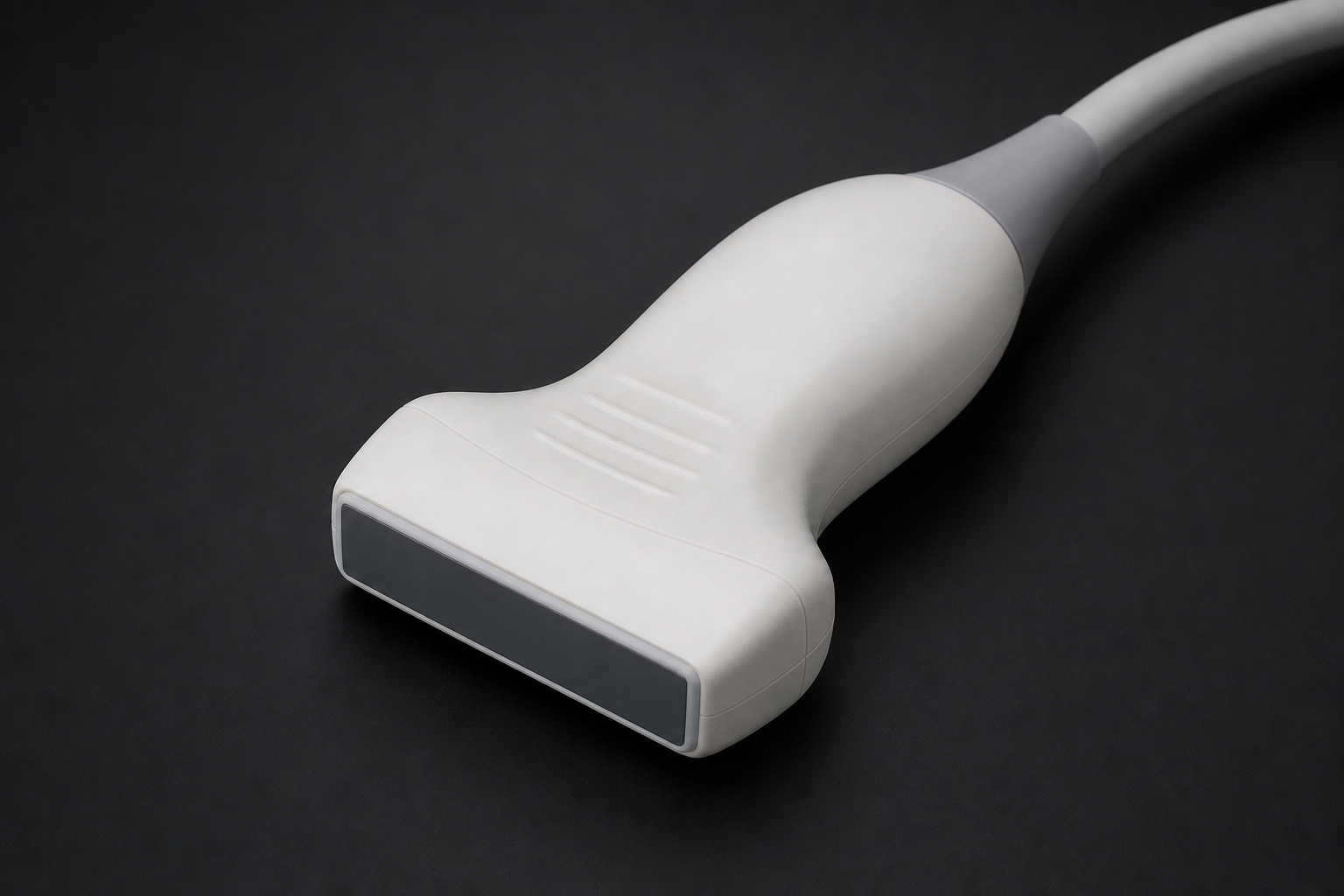
Linear Array
- Shape
- Rectangular
- Frequency
- 5-15 MHz
- Use
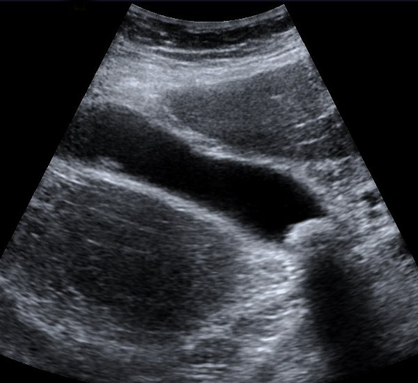
- Vascular, MSK, thyroid, breast
Real-time, safe, and portable — ultrasound uses high-frequency sound waves (1–20 MHz) to produce images of soft tissue structures. No ionizing radiation, no magnets, no contrast agents required for most applications.
Ultrasound uses mechanical sound waves above the human hearing threshold (>20,000 Hz). Diagnostic ultrasound operates at 1–20 MHz. These waves are mechanical pressure waves — they require a medium to travel through (unlike X-rays or light).
The pulse-echo principle: A short ultrasound pulse is transmitted into the body. When it hits a tissue interface (boundary between different tissue types), some energy is reflected back as an echo. The transducer detects the returning echo. The time taken for the echo to return determines the depth of the interface: depth = (speed × time) / 2.
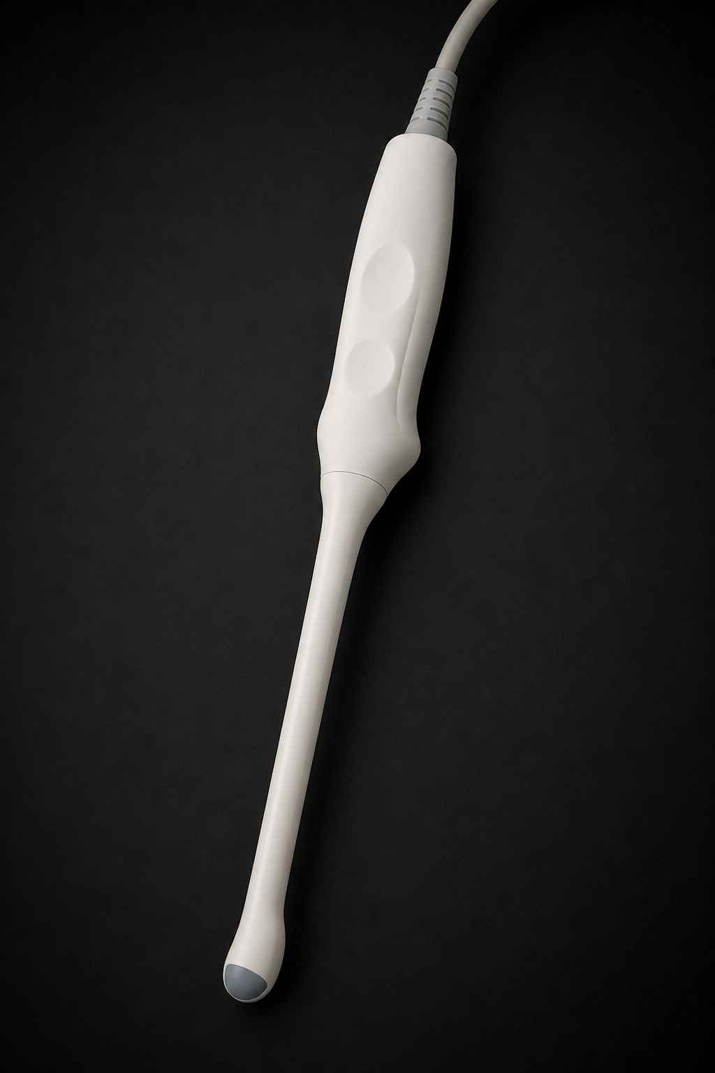
Click any label to explore each component.
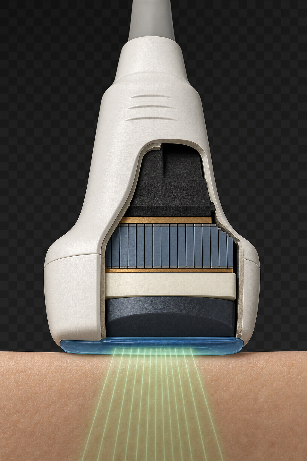
The heart of the ultrasound transducer is the piezoelectric crystal. This material has a remarkable dual property:
Click on a component in the diagram to learn about each layer of the transducer.
Click a mode to see its waveform display and learn how it works.
Amplitude mode — earliest form. Shows echo amplitude vs. depth as a 1D trace.
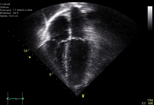
Brightness mode — standard 2D imaging. Echo intensity displayed as pixel brightness.
Motion mode — time plotted against depth. Used for cardiac valve and fetal heart rate measurements.
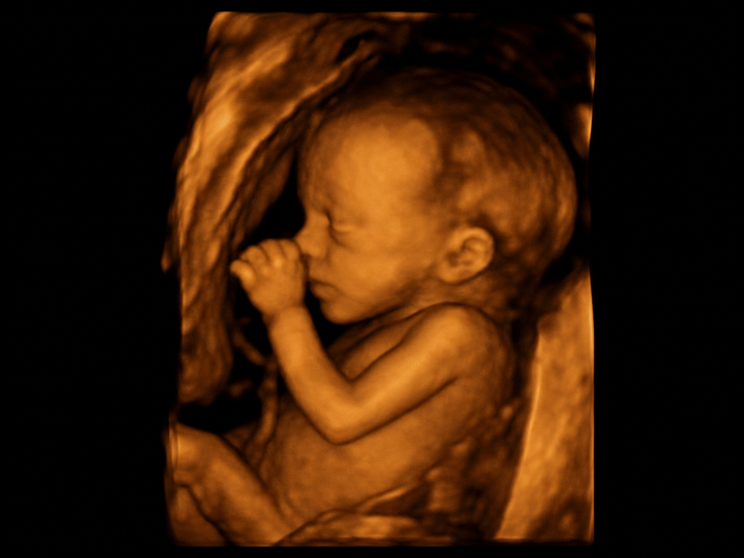
Volumetric imaging. 4D adds real-time motion — popular in obstetric imaging for fetal anatomy.
A-mode (Amplitude) displays echo amplitudes as vertical spikes along a horizontal baseline representing depth. It was the original form of diagnostic ultrasound used in the 1950s. Today it is mainly used in ophthalmology for precise eye measurements.
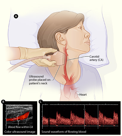
When the sound-reflecting target (e.g., red blood cells) is moving relative to the transducer, the reflected frequency is shifted. This is the Doppler effect. The frequency shift is directly proportional to the velocity of the moving target.
Doppler Equation: Δf = 2 · f₀ · v · cosθ / c
Ultrasound is the safest major imaging modality — preferred in pregnancy, pediatrics, and any situation where ionizing radiation should be avoided.
Safety profile: No known harmful biological effects at diagnostic intensities. Preferred modality during pregnancy and for children. ALARA still applies — minimize unnecessary scanning time and power output.
Image sources: Wikimedia Commons examples for obstetric ultrasound, echocardiography, abdominal ultrasound, vascular ultrasound, musculoskeletal ultrasound, and lung POCUS.
MRI — the most powerful soft-tissue imaging modality, using magnetic fields and radio waves.
Explore MRI →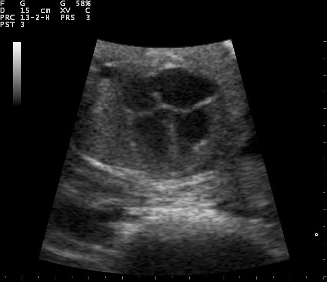
